3L45

A Joint Neutron and X-ray structure of Oxidized Amicyanin


Experimental Data Snapshot

  • Method: NEUTRON DIFFRACTION
  • Resolution: 1.80 Å
  • R-Value Free: 0.234 
  • R-Value Work: 0.209 
  • R-Value Observed: 0.209 

  • Method: X-RAY DIFFRACTION
  • Resolution: 1.50 Å
  • R-Value Free: 0.215 
  • R-Value Work: 0.198 

wwPDB Validation   3D Report Full Report


This is version 1.3 of the entry. See complete history


Literature

A joint x-ray and neutron study on amicyanin reveals the role of protein dynamics in electron transfer.

Sukumar, N.Mathews, F.S.Langan, P.Davidson, V.L.

(2010) Proc Natl Acad Sci U S A 107: 6817-6822

  • DOI: https://doi.org/10.1073/pnas.0912672107
  • Primary Citation of Related Structures:  
    3L45

  • PubMed Abstract: 

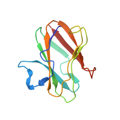
    The joint x-ray/neutron diffraction model of the Type I copper protein, amicyanin from Paracoccus denitrificans was determined at 1.8 A resolution. The protein was crystallized using reagents prepared in D(2)O. About 86% of the amide hydrogen atoms are either partially or fully exchanged, which correlates well with the atomic depth of the amide nitrogen atom and the secondary structure type, but with notable exceptions. Each of the four residues that provide copper ligands is partially deuterated. The model reveals the dynamic nature of the protein, especially around the copper-binding site. A detailed analysis of the presence of deuterated water molecules near the exchange sites indicates that amide hydrogen exchange is primarily due to the flexibility of the protein. Analysis of the electron transfer path through the protein shows that residues in that region are highly dynamic, as judged by hydrogen/deuterium exchange. This could increase the rate of electron transfer by transiently shortening through-space jumps in pathways or by increasing the atomic packing density. Analysis of C-HX bonding reveals previously undefined roles of these relatively weak H bonds, which, when present in sufficient number can collectively influence the structure, redox, and electron transfer properties of amicyanin.


  • Organizational Affiliation

    Northeastern Collaborative Access Team and Department of Chemistry and Chemical Biology, Cornell University, Building 436E, Argonne National Laboratory, Argonne, IL 60439, USA. sukumar@anl.gov


Macromolecules
Find similar proteins by:  (by identity cutoff)  |  3D Structure
Entity ID: 1
MoleculeChains Sequence LengthOrganismDetailsImage
Amicyanin105Paracoccus denitrificansMutation(s): 0 
Gene Names: amimauC
UniProt
Find proteins for P22364 (Paracoccus denitrificans)
Explore P22364 
Go to UniProtKB:  P22364
Entity Groups  
Sequence Clusters30% Identity50% Identity70% Identity90% Identity95% Identity100% Identity
UniProt GroupP22364
Sequence Annotations
Expand
  • Reference Sequence
Small Molecules
Ligands 1 Unique
IDChains Name / Formula / InChI Key2D Diagram3D Interactions
CU
Query on CU

Download Ideal Coordinates CCD File 
B [auth A]COPPER (II) ION
Cu
JPVYNHNXODAKFH-UHFFFAOYSA-N
Experimental Data & Validation

Experimental Data

  • Method: NEUTRON DIFFRACTION
  • Resolution: 1.80 Å
  • R-Value Free: 0.234 
  • R-Value Work: 0.209 
  • R-Value Observed: 0.209 
  • Space Group: P 1 21 1
  • Method: X-RAY DIFFRACTION
  • Resolution: 1.50 Å
  • R-Value Free: 0.215 
  • R-Value Work: 0.198 
  • Space Group: P 1 21 1
Unit Cell:
Length ( Å )Angle ( ˚ )
a = 27.54α = 90
b = 56.58β = 96.21
c = 28.86γ = 90
Software Package:
Software NamePurpose
nCNSrefinement
HKL-2000data reduction
d*TREKdata reduction
HKL-2000data scaling
LAUENORMdata scaling
SCALAdata scaling
EPMRphasing

Structure Validation

View Full Validation Report


Entry History 

Deposition Data

Revision History  (Full details and data files)

  • Version 1.0: 2010-04-28
    Type: Initial release
  • Version 1.1: 2011-07-13
    Changes: Data collection, Version format compliance
  • Version 1.2: 2018-04-25
    Changes: Data collection
  • Version 1.3: 2023-09-13
    Changes: Data collection, Database references, Derived calculations, Refinement description