3KJ4

Structure of rat Nogo receptor bound to 1D9 antagonist antibody


Experimental Data Snapshot

  • Method: X-RAY DIFFRACTION
  • Resolution: 3.10 Å
  • R-Value Free: 0.318 
  • R-Value Work: 0.253 
  • R-Value Observed: 0.256 

wwPDB Validation   3D Report Full Report


Ligand Structure Quality Assessment 


This is version 2.1 of the entry. See complete history


Literature

Resolution of disulfide heterogeneity in Nogo receptor 1 fusion proteins by molecular engineering.

Weinreb, P.H.Wen, D.Qian, F.Wildes, C.P.Garber, E.A.Walus, L.Jung, M.Y.Wang, J.Relton, J.K.Amatucci, J.Wang, R.Porreca, F.Silvian, L.Meier, W.Pepinsky, R.B.Lee, D.H.

(2010) Biotechnol Appl Biochem 57: 31-45

  • DOI: https://doi.org/10.1042/BA20100061
  • Primary Citation of Related Structures:  
    3KJ4

  • PubMed Abstract: 

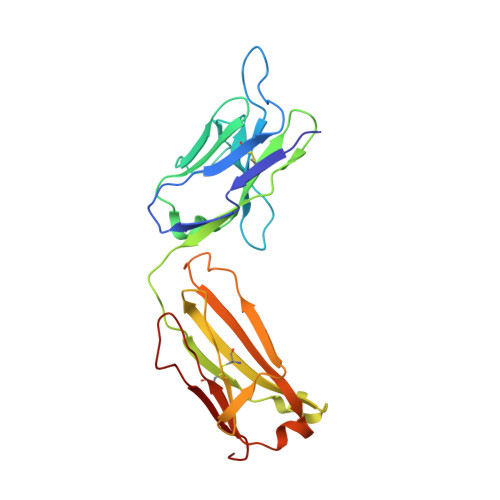
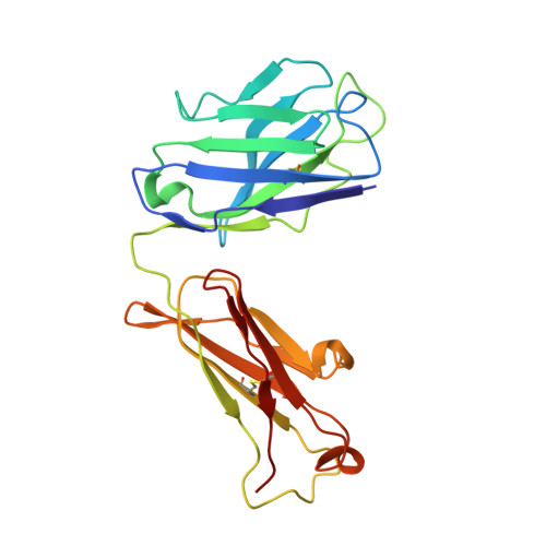
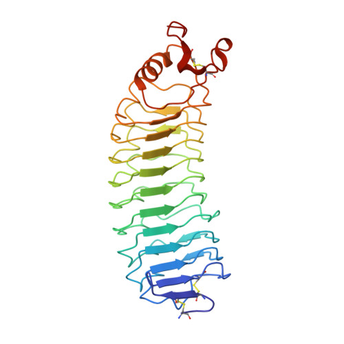
    NgRI (Nogo-66 receptor) is part of a signalling complex that inhibits axon regeneration in the central nervous system. Truncated soluble versions of NgRI have been used successfully to promote axon regeneration in animal models of spinal-cord injury, raising interest in this protein as a potential therapeutic target. The LRR (leucine-rich repeat) regions in NgRI are flanked by N- and C-terminal disulfide-containing 'cap' domains (LRRNT and LRRCT respectively). In the present work we show that, although functionally active, the NgRI(310)-Fc fusion protein contains mislinked and heterogeneous disulfide patterns in the LRRCT domain, and we report the generation of a series of variant molecules specifically designed to prevent this heterogeneity. Using these variants we explored the effects of modifying the NgRI truncation site or the spacing between the NgRI and Fc domains, or replacing cysteines within the NgRI or IgG hinge regions. One variant, which incorporates replacements of Cys²⁶⁶ and Cys³⁰⁹ with alanine residues, completely eliminated disulfide scrambling while maintaining functional in vitro and in vivo efficacy. This modified NgRI-Fc molecule represents a significantly improved candidate for further pharmaceutical development, and may serve as a useful model for the optimization of other IgG fusion proteins made from LRR proteins.


  • Organizational Affiliation

    Biogen Idec, Inc., 14 Cambridge Center, Cambridge, MA 02142, USA. paul.weinreb@biogenidec.com


Macromolecules
Find similar proteins by:  (by identity cutoff)  |  3D Structure
Entity ID: 1
MoleculeChains Sequence LengthOrganismDetailsImage
Fab fragment 1D9 light chainA [auth L],
D [auth B]
219Mus musculusMutation(s): 0 
Entity Groups  
Sequence Clusters30% Identity50% Identity70% Identity90% Identity95% Identity100% Identity
Sequence Annotations
Expand
  • Reference Sequence
Find similar proteins by:  (by identity cutoff)  |  3D Structure
Entity ID: 2
MoleculeChains Sequence LengthOrganismDetailsImage
Fab fragment 1D9 heavy chainB [auth H],
E [auth C]
220Mus musculusMutation(s): 0 
Entity Groups  
Sequence Clusters30% Identity50% Identity70% Identity90% Identity95% Identity100% Identity
Sequence Annotations
Expand
  • Reference Sequence
Find similar proteins by:  (by identity cutoff)  |  3D Structure
Entity ID: 3
MoleculeChains Sequence LengthOrganismDetailsImage
Reticulon-4 receptorC [auth A],
F [auth D]
286Rattus norvegicusMutation(s): 0 
Gene Names: Nogo ReceptorNogorRtn4r
UniProt
Find proteins for Q99M75 (Rattus norvegicus)
Explore Q99M75 
Go to UniProtKB:  Q99M75
Entity Groups  
Sequence Clusters30% Identity50% Identity70% Identity90% Identity95% Identity100% Identity
UniProt GroupQ99M75
Sequence Annotations
Expand
  • Reference Sequence
Small Molecules
Ligands 4 Unique
IDChains Name / Formula / InChI Key2D Diagram3D Interactions
NAG
Query on NAG

Download Ideal Coordinates CCD File 
K [auth A],
O [auth D]
2-acetamido-2-deoxy-beta-D-glucopyranose
C8 H15 N O6
OVRNDRQMDRJTHS-FMDGEEDCSA-N
NDG
Query on NDG

Download Ideal Coordinates CCD File 
I [auth A],
J [auth A],
M [auth D],
N [auth D]
2-acetamido-2-deoxy-alpha-D-glucopyranose
C8 H15 N O6
OVRNDRQMDRJTHS-PVFLNQBWSA-N
MAN
Query on MAN

Download Ideal Coordinates CCD File 
G [auth H]alpha-D-mannopyranose
C6 H12 O6
WQZGKKKJIJFFOK-PQMKYFCFSA-N
ZN
Query on ZN

Download Ideal Coordinates CCD File 
H,
L [auth C]
ZINC ION
Zn
PTFCDOFLOPIGGS-UHFFFAOYSA-N
Experimental Data & Validation

Experimental Data

  • Method: X-RAY DIFFRACTION
  • Resolution: 3.10 Å
  • R-Value Free: 0.318 
  • R-Value Work: 0.253 
  • R-Value Observed: 0.256 
  • Space Group: P 21 21 2
Unit Cell:
Length ( Å )Angle ( ˚ )
a = 188.664α = 90
b = 125.488β = 90
c = 90.575γ = 90
Software Package:
Software NamePurpose
ADSCdata collection
PHASERphasing
REFMACrefinement
DENZOdata reduction
SCALEPACKdata scaling

Structure Validation

View Full Validation Report


Ligand Structure Quality Assessment 


Entry History 

Deposition Data

Revision History  (Full details and data files)

  • Version 1.0: 2010-09-22
    Type: Initial release
  • Version 1.1: 2011-07-13
    Changes: Version format compliance
  • Version 2.0: 2020-07-29
    Type: Remediation
    Reason: Carbohydrate remediation
    Changes: Advisory, Atomic model, Data collection, Derived calculations, Structure summary
  • Version 2.1: 2023-09-06
    Changes: Data collection, Database references, Refinement description, Structure summary