Specific Inhibition of the Aspartate Aminotransferase of Plasmodium falciparum.
Wrenger, C., Muller, I.B., Schifferdecker, A.J., Jain, R., Jordanova, R., Groves, M.R.(2011) J Mol Biology 405: 956-971
- PubMed: 21087616
- DOI: https://doi.org/10.1016/j.jmb.2010.11.018
- Primary Citation of Related Structures:
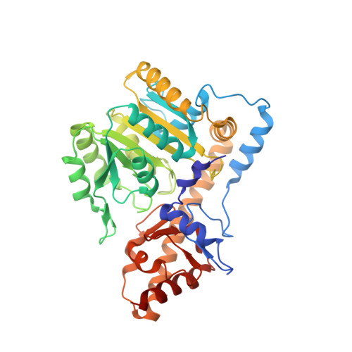
3K7Y - PubMed Abstract:
Aspartate aminotransferases (AspATs; EC 2.6.1.1) catalyze the conversion of aspartate and α-ketoglutarate into oxaloacetate and glutamate and are key enzymes in the nitrogen metabolism of all organisms. Recent findings suggest that the plasmodial enzyme [Plasmodium falciparum aspartate aminotransferase (PfAspAT)] may also play a pivotal role in energy metabolism and in the de novo biosynthesis of pyrimidines. However, while PfAspAT is a potential drug target, the high homology between the active sites of currently available AspAT structures hinders the development of specific inhibitors of these enzymes. In this article, we report the X-ray structure of the PfAspAT homodimer at a resolution of 2.8 Å. While the overall fold is similar to the currently available structures of other AspATs, the structure presented shows a significant divergence in the conformation of the N-terminal residues. Deletion of these divergent PfAspAT N-terminal residues results in a loss of activity for the recombinant protein, and addition of a peptide containing these 13 N-terminal residues results in inhibition both in vitro and in a lysate isolated from cultured parasites, while the activity of human cytosolic AspAT is unaffected. The finding that the divergent N-terminal amino acids of PfAspAT play a role in catalytic activity indicates that specific inhibition of the enzyme may provide a lead for the development of novel compounds in the treatment of malaria. We also report on the localization of PfAspAT to the parasite cytosol and discuss the implications of the role of PfAspAT in the supply of malate to the parasite mitochondria.
- Department of Biochemistry, Bernhard Nocht Institute for Tropical Medicine, Bernhard Nocht Strasse 74, D-20359 Hamburg, Germany.
Organizational Affiliation: