3ERX

High-resolution structure of Paracoccus pantotrophus pseudoazurin


Experimental Data Snapshot

  • Method: X-RAY DIFFRACTION
  • Resolution: 1.25 Å
  • R-Value Free: 0.206 
  • R-Value Work: 0.188 
  • R-Value Observed: 0.189 

wwPDB Validation   3D Report Full Report


This is version 1.2 of the entry. See complete history


Literature

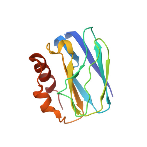
The 1.4 A resolution structure of Paracoccus pantotrophus pseudoazurin.

Najmudin, S.Pauleta, S.R.Moura, I.Romao, M.J.

(2010) Acta Crystallogr Sect F Struct Biol Cryst Commun 66: 627-635

  • DOI: https://doi.org/10.1107/S1744309110013989
  • Primary Citation of Related Structures:  
    3ERX

  • PubMed Abstract: 

    Pseudoazurins are small type 1 copper proteins that are involved in the flow of electrons between various electron donors and acceptors in the bacterial periplasm, mostly under denitrifying conditions. The previously determined structure of Paracoccus pantotrophus pseudoazurin in the oxidized form was improved to a nominal resolution of 1.4 A, with R and R(free) values of 0.188 and 0.206, respectively. This high-resolution structure makes it possible to analyze the interactions between the monomers and the solvent structure in detail. Analysis of the high-resolution structure revealed the structural regions that are responsible for monomer-monomer recognition during dimer formation and for protein-protein interaction and that are important for partner recognition. The pseudoazurin structure was compared with other structures of various type 1 copper proteins and these were grouped into families according to similarities in their secondary structure; this may be useful in the annotation of copper proteins in newly sequenced genomes and in the identification of novel copper proteins.


  • Organizational Affiliation

    REQUIMTE, Centro de Química Fina e Biotecnologia, Departamento de Química, Faculdade de Ciências e Tecnologia, Universidade Nova de Lisboa, 2829-516 Caparica, Portugal. shabir@fmv.utl.pt


Macromolecules
Find similar proteins by:  (by identity cutoff)  |  3D Structure
Entity ID: 1
MoleculeChains Sequence LengthOrganismDetailsImage
Pseudoazurin
A, B
123Paracoccus pantotrophusMutation(s): 0 
Gene Names: pazS
UniProt
Find proteins for P80401 (Paracoccus pantotrophus)
Explore P80401 
Go to UniProtKB:  P80401
Entity Groups  
Sequence Clusters30% Identity50% Identity70% Identity90% Identity95% Identity100% Identity
UniProt GroupP80401
Sequence Annotations
Expand
  • Reference Sequence
Experimental Data & Validation

Experimental Data

  • Method: X-RAY DIFFRACTION
  • Resolution: 1.25 Å
  • R-Value Free: 0.206 
  • R-Value Work: 0.188 
  • R-Value Observed: 0.189 
  • Space Group: C 1 2 1
Unit Cell:
Length ( Å )Angle ( ˚ )
a = 108.823α = 90
b = 56.993β = 126.22
c = 67.982γ = 90
Software Package:
Software NamePurpose
ADSCdata collection
PHASERphasing
REFMACrefinement
MOSFLMdata reduction
SCALAdata scaling

Structure Validation

View Full Validation Report


Entry History 

Deposition Data

Revision History  (Full details and data files)

  • Version 1.0: 2009-10-13
    Type: Initial release
  • Version 1.1: 2011-07-13
    Changes: Advisory, Version format compliance
  • Version 1.2: 2023-09-06
    Changes: Data collection, Database references, Derived calculations, Refinement description