3DPC

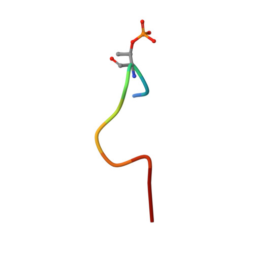
Structure of E.coli Alkaline Phosphatase Mutant in Complex with a Phosphorylated Peptide


Experimental Data Snapshot

  • Method: X-RAY DIFFRACTION
  • Resolution: 2.30 Å
  • R-Value Free: 0.271 
  • R-Value Work: 0.234 

wwPDB Validation   3D Report Full Report


This is version 1.4 of the entry. See complete history


Literature

Development of a universal phosphorylated peptide-binding protein for simultaneous assay of kinases

Li, W.Bi, L.Wang, W.Li, Y.Zhou, Y.Wei, H.Jiang, T.Bai, L.Chen, Y.Zhang, Z.Yuan, X.Xiao, J.Zhang, X.-E.

(2009) Biosens Bioelectron 24: 2871-2877

  • DOI: https://doi.org/10.1016/j.bios.2009.02.020
  • Primary Citation of Related Structures:  
    3DPC

  • PubMed Abstract: 

    This study describes the development of a universal phosphorylated peptide-binding protein designed to simultaneously detect serine, threonine and tyrosine kinases. The Escherichia coli alkaline phosphatase (EAP) is a well-defined nonspecific phosphated monoesterase and Ser-, Thr- or Tyr-phosphorylated peptides served as substrates for EAP in preliminary experiments. Based on the known catalytic mechanism of EAP, the recombinant site-directed mutant EAP-S102L was generated, whose catalytic activity was blocked, but its binding ability was preserved. For EAP-S102L the catalytic rate constant, k(cat), was reduced by a factor of 1000, while the Michaelis-Menten constant, K(m), remained almost unchanged. Crystallographic analysis of the EAP-S102L/phophorylated peptide complex revealed that EAP-S102L could bind the phosphate group of the phosphorylated peptide but lacked nucleophilic attack potential which was essential for the catalytic ability of EAP. Finally, by combining the fluorescence-labeled EAP-S102L with non-phophorylated peptide chips, kinases could be detected from tumor cell samples. The recombinant EAP-S102L construct is perhaps the first functional binding protein derived from a native enzyme, illustrating how one single mutation tremendously alters protein function.


  • Organizational Affiliation

    State Key Laboratory of Virology, Wuhan Institute of Virology, Chinese Academy of Sciences, Wuhan 430071, China.


Macromolecules
Find similar proteins by:  (by identity cutoff)  |  3D Structure
Entity ID: 1
MoleculeChains Sequence LengthOrganismDetailsImage
Alkaline phosphatase
A, B
455Escherichia coli K-12Mutation(s): 1 
Gene Names: phoAb0383JW0374
EC: 3.1.3.1
UniProt
Find proteins for P00634 (Escherichia coli (strain K12))
Explore P00634 
Go to UniProtKB:  P00634
Entity Groups  
Sequence Clusters30% Identity50% Identity70% Identity90% Identity95% Identity100% Identity
UniProt GroupP00634
Sequence Annotations
Expand
  • Reference Sequence

Find similar proteins by:  Sequence   |   3D Structure  

Entity ID: 2
MoleculeChains Sequence LengthOrganismDetailsImage
Phosphorylated Peptide10N/AMutation(s): 0 
Entity Groups  
Sequence Clusters30% Identity50% Identity70% Identity90% Identity95% Identity100% Identity
Sequence Annotations
Expand
  • Reference Sequence
Small Molecules
Ligands 2 Unique
IDChains Name / Formula / InChI Key2D Diagram3D Interactions
TRS
Query on TRS

Download Ideal Coordinates CCD File 
E [auth A]2-AMINO-2-HYDROXYMETHYL-PROPANE-1,3-DIOL
C4 H12 N O3
LENZDBCJOHFCAS-UHFFFAOYSA-O
PO4
Query on PO4

Download Ideal Coordinates CCD File 
D [auth A]PHOSPHATE ION
O4 P
NBIIXXVUZAFLBC-UHFFFAOYSA-K
Modified Residues  1 Unique
IDChains TypeFormula2D DiagramParent
TPO
Query on TPO
C
L-PEPTIDE LINKINGC4 H10 N O6 PTHR
Experimental Data & Validation

Experimental Data

  • Method: X-RAY DIFFRACTION
  • Resolution: 2.30 Å
  • R-Value Free: 0.271 
  • R-Value Work: 0.234 
  • Space Group: P 21 21 21
Unit Cell:
Length ( Å )Angle ( ˚ )
a = 64.862α = 90
b = 107.74β = 90
c = 148.389γ = 90
Software Package:
Software NamePurpose
CrystalCleardata collection
PHASESphasing
CNSrefinement
d*TREKdata reduction
d*TREKdata scaling

Structure Validation

View Full Validation Report


Entry History 

Deposition Data

Revision History  (Full details and data files)

  • Version 1.0: 2009-06-16
    Type: Initial release
  • Version 1.1: 2011-07-13
    Changes: Source and taxonomy, Version format compliance
  • Version 1.2: 2014-02-05
    Changes: Derived calculations
  • Version 1.3: 2021-11-10
    Changes: Database references, Derived calculations
  • Version 1.4: 2023-11-01
    Changes: Data collection, Refinement description