3B3K

Crystal structure of the complex between PPARgamma and the full agonist LT175


Experimental Data Snapshot

  • Method: X-RAY DIFFRACTION
  • Resolution: 2.60 Å
  • R-Value Free: 0.316 
  • R-Value Work: 0.237 
  • R-Value Observed: 0.245 

wwPDB Validation   3D Report Full Report


Ligand Structure Quality Assessment 


This is version 1.2 of the entry. See complete history


Literature

Crystal Structure of the Peroxisome Proliferator-Activated Receptor gamma (PPARgamma) Ligand Binding Domain Complexed with a Novel Partial Agonist: A New Region of the Hydrophobic Pocket Could Be Exploited for Drug Design

Montanari, R.Saccoccia, F.Scotti, E.Crestani, M.Godio, C.Gilardi, F.Loiodice, F.Fracchiolla, G.Laghezza, A.Tortorella, P.Lavecchia, A.Novellino, E.Mazza, F.Aschi, M.Pochetti, G.

(2008) J Med Chem 51: 7768-7776

  • DOI: https://doi.org/10.1021/jm800733h
  • Primary Citation of Related Structures:  
    3B3K, 3CDS, 3D6D

  • PubMed Abstract: 

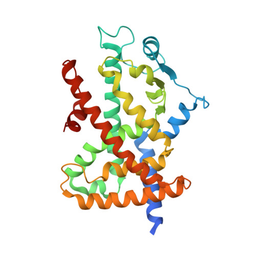
    The peroxisome proliferator-activated receptors (PPARs) are ligand-dependent transcription factors regulating glucose and lipid metabolism. The search for new PPAR ligands with reduced adverse effects with respect to the marketed antidiabetic agents thiazolidinediones (TZDs) and the dual-agonists glitazars is highly desired. We report the crystal structure and activity of the two enantiomeric forms of a clofibric acid analogue, respectively complexed with the ligand-binding domain (LBD) of PPARgamma, and provide an explanation on a molecular basis for their different potency and efficacy against PPARgamma. The more potent S-enantiomer is a dual PPARalpha/PPARgamma agonist which presents a partial agonism profile against PPARgamma. Docking of the S-enantiomer in the PPARalpha-LBD has been performed to explain its different subtype pharmacological profile. The hypothesis that partial agonists show differential stabilization of helix 3, when compared to full agonists, is also discussed. Moreover, the structure of the complex with the S-enantiomer reveals a new region of the PPARgamma-LBD never sampled before by other ligands.


  • Organizational Affiliation

    Consiglio Nazionale delle Ricerche, Roma 00016, Italy.


Macromolecules
Find similar proteins by:  (by identity cutoff)  |  3D Structure
Entity ID: 1
MoleculeChains Sequence LengthOrganismDetailsImage
Peroxisome proliferator-activated receptor gamma
A, B
286Homo sapiensMutation(s): 0 
Gene Names: PPARGNR1C3
UniProt & NIH Common Fund Data Resources
Find proteins for P37231 (Homo sapiens)
Explore P37231 
Go to UniProtKB:  P37231
PHAROS:  P37231
GTEx:  ENSG00000132170 
Entity Groups  
Sequence Clusters30% Identity50% Identity70% Identity90% Identity95% Identity100% Identity
UniProt GroupP37231
Sequence Annotations
Expand
  • Reference Sequence
Small Molecules
Ligands 1 Unique
IDChains Name / Formula / InChI Key2D Diagram3D Interactions
LRG
Query on LRG

Download Ideal Coordinates CCD File 
C [auth A](2S)-2-(biphenyl-4-yloxy)-3-phenylpropanoic acid
C21 H18 O3
TZTPJJNNACUQQR-FQEVSTJZSA-N
Binding Affinity Annotations 
IDSourceBinding Affinity
LRG BindingDB:  3B3K EC50: min: 480, max: 562 (nM) from 5 assay(s)
Experimental Data & Validation

Experimental Data

  • Method: X-RAY DIFFRACTION
  • Resolution: 2.60 Å
  • R-Value Free: 0.316 
  • R-Value Work: 0.237 
  • R-Value Observed: 0.245 
  • Space Group: C 1 2 1
Unit Cell:
Length ( Å )Angle ( ˚ )
a = 93.144α = 90
b = 61.569β = 103.4
c = 118.797γ = 90
Software Package:
Software NamePurpose
MAR345dtbdata collection
CNSrefinement
DENZOdata reduction
SCALEPACKdata scaling
CNSphasing

Structure Validation

View Full Validation Report


Ligand Structure Quality Assessment 


Entry History 

Deposition Data

Revision History  (Full details and data files)

  • Version 1.0: 2008-10-28
    Type: Initial release
  • Version 1.1: 2011-07-13
    Changes: Version format compliance
  • Version 1.2: 2023-11-01
    Changes: Data collection, Database references, Derived calculations, Refinement description