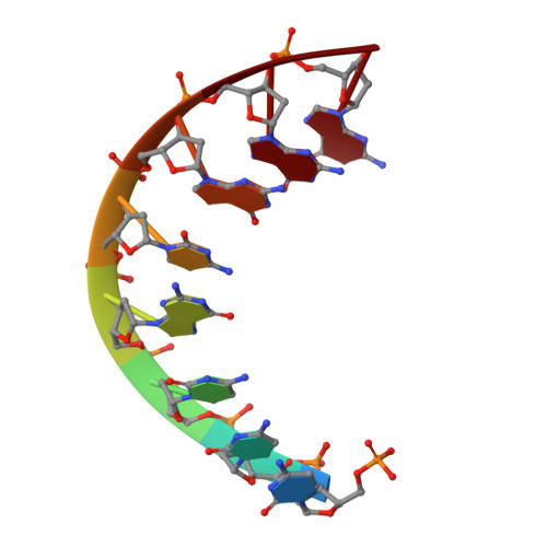
Structural variability of A-DNA in crystals of the octamer d(pCpCpCpGpCpGpGpG)
Fernandez, L.G., Subirana, J.A., Verdaguer, N., Pyshnyi, D., Campos, L., Malinina, L.(1997) J Biomol Struct Dyn 15: 151-163
- PubMed: 9283988
- DOI: https://doi.org/10.1080/07391102.1997.10508954
- Primary Citation of Related Structures:
368D, 369D, 370D, 371D, 372D - PubMed Abstract:
We have determined the structure of the synthetic DNA octamer d(pCpCpCpGpCpGpGpG) in five different crystal forms by single crystal X-ray diffraction. One crystal belongs to the space group P4(3)2(1)2 with a = b = 41.77, c = 25.15 A, whereas all others have the space group P2(1)2(1)2(1) with progressively decreasing unit cell volumes. In all crystals the octamer forms duplexes of A-DNA and all crystals display a similar packing mode, typical for A-DNA. The structure of the duplex varies from loose to very compact when going from one crystal form to another. The most compact form exhibits a volume of 995 A3 per base pair. Such a high density has never been found in A-DNA, being more characteristic of Z-DNA crystals. A comparison of the most with the least compact forms gives a RMS value of 1.7 A, with the distance between the phosphate centers through the major groove being almost twice shorter in the compact form. The phosphate-phosphate separation across the major groove in the compact form is extremely small, 0.7 A. The helical parameters also vary significantly in the various crystal forms. Differences in the helical twist can reach 13 degrees in the same step of the octamer in different crystal forms. The results prove that A-DNA is structurally very variable and demonstrate that the local structure of the same DNA fragment can strongly depend on the crystal environment.
- Engelhardt Institute of Molecular Biology RAS, Moscow, Russia.
Organizational Affiliation: