2UV3

Structure of the signal-regulatory protein (SIRP) alpha domain that binds CD47.


Experimental Data Snapshot

  • Method: X-RAY DIFFRACTION
  • Resolution: 1.80 Å
  • R-Value Free: 0.249 
  • R-Value Work: 0.204 
  • R-Value Observed: 0.207 

wwPDB Validation   3D Report Full Report


This is version 1.2 of the entry. See complete history


Literature

The Structure of the Macrophage Signal Regulatory Protein Alpha (Sirpalpha) Inhibitory Receptor Reveals a Binding Face Reminiscent of that Used by T Cell Receptors.

Hatherley, D.Harlos, K.Dunlop, D.C.Stuart, D.I.Barclay, A.N.

(2007) J Biol Chem 282: 14567

  • DOI: https://doi.org/10.1074/jbc.M611511200
  • Primary Citation of Related Structures:  
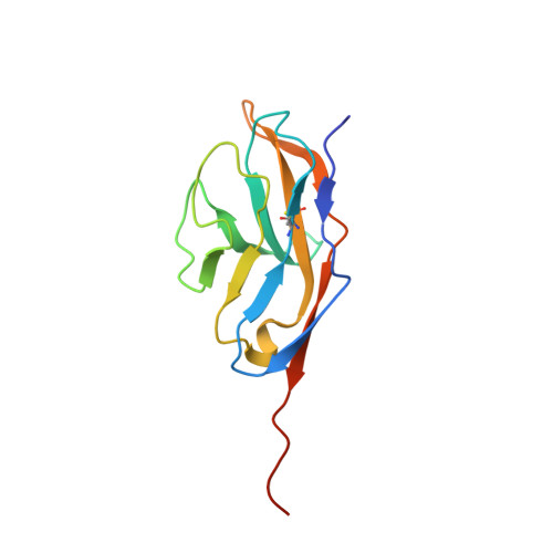
    2UV3

  • PubMed Abstract: 

    Signal regulatory protein (SIRP) alpha is a membrane receptor that sends inhibitory signals to myeloid cells by engagement of CD47. The high resolution x-ray structure of the N-terminal ligand binding domain shows it to have a distinctive immunoglobulin superfamily V-like fold. Site-directed mutagenesis suggests that CD47 is bound at a surface involving the BC, FG, and DE loops, which distinguishes it from other immunoglobulin superfamily surface proteins that use the faces of the fold, but resembles antigen receptors. The SIRP interaction is confined to a single domain, and its use of an extended DE loop strengthens the similarity with T cell receptor binding and the suggestion that they are closely related in evolution. The employment of loops to form the CD47-binding surface provides a mechanism for small sequence changes to modulate binding specificity, explaining the different binding properties of SIRP family members.


  • Organizational Affiliation

    Sir William Dunn School of Pathology, University of Oxford, Oxford, UK.


Macromolecules
Find similar proteins by:  (by identity cutoff)  |  3D Structure
Entity ID: 1
MoleculeChains Sequence LengthOrganismDetailsImage
TYROSINE-PROTEIN PHOSPHATASE NON-RECEPTOR TYPE SUBSTRATE 1
A, B
126Homo sapiensMutation(s): 0 
UniProt & NIH Common Fund Data Resources
Find proteins for P78324 (Homo sapiens)
Explore P78324 
Go to UniProtKB:  P78324
PHAROS:  P78324
GTEx:  ENSG00000198053 
Entity Groups  
Sequence Clusters30% Identity50% Identity70% Identity90% Identity95% Identity100% Identity
UniProt GroupP78324
Sequence Annotations
Expand
  • Reference Sequence
Experimental Data & Validation

Experimental Data

  • Method: X-RAY DIFFRACTION
  • Resolution: 1.80 Å
  • R-Value Free: 0.249 
  • R-Value Work: 0.204 
  • R-Value Observed: 0.207 
  • Space Group: C 1 2 1
Unit Cell:
Length ( Å )Angle ( ˚ )
a = 76.831α = 90
b = 56.266β = 114.9
c = 82.245γ = 90
Software Package:
Software NamePurpose
REFMACrefinement
HKL-2000data reduction
SCALEPACKdata scaling
PHASERphasing

Structure Validation

View Full Validation Report


Entry History 

Deposition Data

Revision History  (Full details and data files)

  • Version 1.0: 2007-03-27
    Type: Initial release
  • Version 1.1: 2011-07-13
    Changes: Advisory, Refinement description, Version format compliance
  • Version 1.2: 2024-05-01
    Changes: Data collection, Database references, Other, Refinement description