2QW7

Carboxysome Subunit, CcmL


Experimental Data Snapshot

  • Method: X-RAY DIFFRACTION
  • Resolution: 2.40 Å
  • R-Value Free: 0.296 
  • R-Value Work: 0.240 
  • R-Value Observed: 0.243 

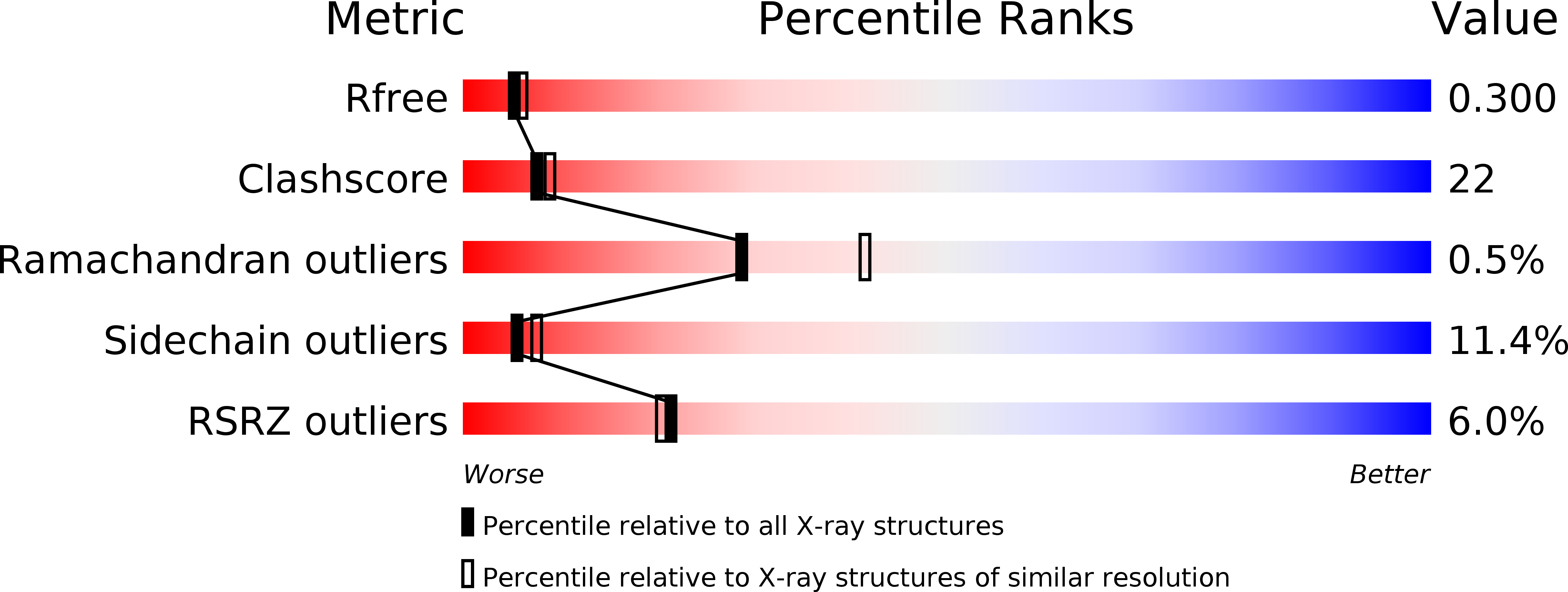
wwPDB Validation   3D Report Full Report


This is version 1.2 of the entry. See complete history


Literature

Atomic-level models of the bacterial carboxysome shell.

Tanaka, S.Kerfeld, C.A.Sawaya, M.R.Cai, F.Heinhorst, S.Cannon, G.C.Yeates, T.O.

(2008) Science 319: 1083-1086

  • DOI: https://doi.org/10.1126/science.1151458
  • Primary Citation of Related Structures:  
    2QW7, 2RCF, 3BN4

  • PubMed Abstract: 

    The carboxysome is a bacterial microcompartment that functions as a simple organelle by sequestering enzymes involved in carbon fixation. The carboxysome shell is roughly 800 to 1400 angstroms in diameter and is assembled from several thousand protein subunits. Previous studies have revealed the three-dimensional structures of hexameric carboxysome shell proteins, which self-assemble into molecular layers that most likely constitute the facets of the polyhedral shell. Here, we report the three-dimensional structures of two proteins of previously unknown function, CcmL and OrfA (or CsoS4A), from the two known classes of carboxysomes, at resolutions of 2.4 and 2.15 angstroms. Both proteins assemble to form pentameric structures whose size and shape are compatible with formation of vertices in an icosahedral shell. Combining these pentamers with the hexamers previously elucidated gives two plausible, preliminary atomic models for the carboxysome shell.


  • Organizational Affiliation

    Department of Chemistry and Biochemistry, University of California at Los Angeles (UCLA), Los Angeles, CA 90095, USA.


Macromolecules
Find similar proteins by:  (by identity cutoff)  |  3D Structure
Entity ID: 1
MoleculeChains Sequence LengthOrganismDetailsImage
Carbon dioxide concentrating mechanism protein ccmL
A, B, C, D, E
A, B, C, D, E, F, G, H, I, J
111Synechocystis sp. PCC 6803Mutation(s): 0 
Gene Names: ccmL
UniProt
Find proteins for P72759 (Synechocystis sp. (strain PCC 6803 / Kazusa))
Explore P72759 
Go to UniProtKB:  P72759
Entity Groups  
Sequence Clusters30% Identity50% Identity70% Identity90% Identity95% Identity100% Identity
UniProt GroupP72759
Sequence Annotations
Expand
  • Reference Sequence
Experimental Data & Validation

Experimental Data

  • Method: X-RAY DIFFRACTION
  • Resolution: 2.40 Å
  • R-Value Free: 0.296 
  • R-Value Work: 0.240 
  • R-Value Observed: 0.243 
  • Space Group: P 1 21 1
Unit Cell:
Length ( Å )Angle ( ˚ )
a = 64.428α = 90
b = 107.444β = 96.51
c = 72.264γ = 90
Software Package:
Software NamePurpose
REFMACrefinement
PDB_EXTRACTdata extraction

Structure Validation

View Full Validation Report


Entry History 

Deposition Data

Revision History  (Full details and data files)

  • Version 1.0: 2008-03-04
    Type: Initial release
  • Version 1.1: 2011-07-13
    Changes: Advisory, Source and taxonomy, Version format compliance
  • Version 1.2: 2024-02-21
    Changes: Data collection, Database references, Derived calculations, Refinement description