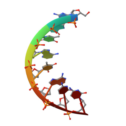
2PLB

D(GTATACC) under hydrostatic pressure of 1.39 GPa


Experimental Data Snapshot

  • Method: X-RAY DIFFRACTION
  • Resolution: 1.60 Å
  • R-Value Free: 0.222 
  • R-Value Work: 0.181 
  • R-Value Observed: 0.187 

wwPDB Validation   3D Report Full Report


Ligand Structure Quality Assessment 


This is version 1.3 of the entry. See complete history


Literature

Adaptation of the base-paired double-helix molecular architecture to extreme pressure.

Girard, E.Prange, T.Dhaussy, A.C.Migianu-Griffoni, E.Lecouvey, M.Chervin, J.C.Mezouar, M.Kahn, R.Fourme, R.

(2007) Nucleic Acids Res 35: 4800-4808

  • DOI: https://doi.org/10.1093/nar/gkm511
  • Primary Citation of Related Structures:  
    2PKV, 2PL4, 2PL8, 2PLB

  • PubMed Abstract: 

    The behaviour of the d(GGTATACC) oligonucleotide has been investigated by X-ray crystallography at 295 K in the range from ambient pressure to 2 GPa (approximately 20,000 atm). Four 3D-structures of the A-DNA form (at ambient pressure, 0.55, 1.09 and 1.39 GPa) were refined at 1.60 or 1.65 A resolution. In addition to the diffraction pattern of the A-form, the broad meridional streaks previously explained by occluded B-DNA octamers within the channels of the crystalline A-form matrix were observed up to at least 2 GPa. This work highlights an important property of nucleic acids, their capability to withstand very high pressures, while keeping in such conditions a nearly invariant geometry of base pairs that store and carry genetic information. The double-helix base-paired architecture behaves as a molecular spring, which makes it especially adapted to very harsh conditions. These features may have contributed to the emergence of a RNA World at prebiotic stage.


  • Organizational Affiliation

    Synchrotron-SOLEIL, L'Orme des Merisiers, Saint-Aubin, BP 48, 91192 Gif-sur-Yvette Cedex, France. eric.girard@synchrotron-soleil.fr


Macromolecules

Find similar nucleic acids by:  Sequence   |   3D Structure  

Entity ID: 1
MoleculeChains LengthOrganismImage
5'-D(*DGP*DGP*DTP*DAP*DTP*DAP*DCP*DC)-3'
A, B
8N/A
Sequence Annotations
Expand
  • Reference Sequence
Small Molecules
Ligands 1 Unique
IDChains Name / Formula / InChI Key2D Diagram3D Interactions
SPM
Query on SPM

Download Ideal Coordinates CCD File 
C [auth B]SPERMINE
C10 H26 N4
PFNFFQXMRSDOHW-UHFFFAOYSA-N
Experimental Data & Validation

Experimental Data

  • Method: X-RAY DIFFRACTION
  • Resolution: 1.60 Å
  • R-Value Free: 0.222 
  • R-Value Work: 0.181 
  • R-Value Observed: 0.187 
  • Space Group: P 61
Unit Cell:
Length ( Å )Angle ( ˚ )
a = 42.83α = 90
b = 42.83β = 90
c = 40.3γ = 120
Software Package:
Software NamePurpose
SHELXmodel building
SHELXL-97refinement
AUTOMARdata collection
XDSdata reduction
SCALAdata scaling

Structure Validation

View Full Validation Report


Ligand Structure Quality Assessment 


Entry History 

Deposition Data

Revision History  (Full details and data files)

  • Version 1.0: 2007-07-24
    Type: Initial release
  • Version 1.1: 2008-01-17
    Changes: Version format compliance
  • Version 1.2: 2011-07-13
    Changes: Version format compliance
  • Version 1.3: 2023-08-30
    Changes: Data collection, Database references, Derived calculations, Refinement description