2PE5

Crystal Structure of the Lac Repressor bound to ONPG in repressed state


Experimental Data Snapshot

  • Method: X-RAY DIFFRACTION
  • Resolution: 3.50 Å
  • R-Value Free: 0.287 
  • R-Value Work: 0.233 
  • R-Value Observed: 0.236 

wwPDB Validation   3D Report Full Report


Ligand Structure Quality Assessment 


This is version 2.2 of the entry. See complete history


Literature

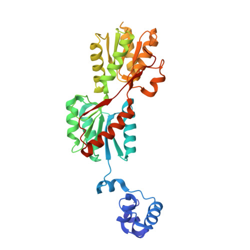
Structural analysis of lac repressor bound to allosteric effectors

Daber, R.Stayrook, S.Rosenberg, A.Lewis, M.

(2007) J Mol Biol 370: 609-619

  • DOI: https://doi.org/10.1016/j.jmb.2007.04.028
  • Primary Citation of Related Structures:  
    2P9H, 2PAF, 2PE5

  • PubMed Abstract: 

    The lac operon is a model system for understanding how effector molecules regulate transcription and are necessary for allosteric transitions. The crystal structures of the lac repressor bound to inducer and anti-inducer molecules provide a model for how these small molecules can modulate repressor function. The structures of the apo repressor and the repressor bound to effector molecules are compared in atomic detail. All effectors examined here bind to the repressor in the same location and are anchored to the repressor through hydrogen bonds to several hydroxyl groups of the sugar ring. Inducer molecules form a more extensive hydrogen-bonding network compared to anti-inducers and neutral effector molecules. The structures of these effector molecules suggest that the O6 hydroxyl on the galactoside is essential for establishing a water-mediated hydrogen bonding network that bridges the N-terminal and C-terminal sub-domains. The altered hydrogen bonding can account in part for the different structural conformations of the repressor, and is vital for the allosteric transition.


  • Organizational Affiliation

    Department of Biochemistry and Biophysics, University of Pennsylvania School of Medicine, 37th and Hamilton Walk, Philadelphia, PA 19104-6059, USA.


Macromolecules

Find similar proteins by:  (by identity cutoff)  |  3D Structure
Entity ID: 2
MoleculeChains Sequence LengthOrganismDetailsImage
Lactose operon repressorD [auth A],
E [auth B],
F [auth C]
330Escherichia coliMutation(s): 1 
Gene Names: lacI
UniProt
Find proteins for P03023 (Escherichia coli (strain K12))
Explore P03023 
Go to UniProtKB:  P03023
Entity Groups  
Sequence Clusters30% Identity50% Identity70% Identity90% Identity95% Identity100% Identity
UniProt GroupP03023
Sequence Annotations
Expand
  • Reference Sequence

Find similar nucleic acids by:  Sequence   |   3D Structure  

Entity ID: 1
MoleculeChains LengthOrganismImage
DNA (5'-D(*DAP*DAP*DTP*DTP*DGP*DTP*DGP*DAP*DGP*DCP*DGP*DCP*DTP*DCP*DAP*DCP*DAP*DAP*DTP*DT)-3')A [auth D],
B [auth E],
C [auth F]
20synthetic construct
Sequence Annotations
Expand
  • Reference Sequence
Experimental Data & Validation

Experimental Data

  • Method: X-RAY DIFFRACTION
  • Resolution: 3.50 Å
  • R-Value Free: 0.287 
  • R-Value Work: 0.233 
  • R-Value Observed: 0.236 
  • Space Group: H 3 2
Unit Cell:
Length ( Å )Angle ( ˚ )
a = 253.08α = 90
b = 253.08β = 90
c = 203.247γ = 120
Software Package:
Software NamePurpose
REFMACrefinement
MAR345data collection
DENZOdata reduction
SCALEPACKdata scaling
PHASERphasing

Structure Validation

View Full Validation Report


Ligand Structure Quality Assessment 


Entry History 

Deposition Data

Revision History  (Full details and data files)

  • Version 1.0: 2008-03-18
    Type: Initial release
  • Version 1.1: 2011-07-13
    Changes: Version format compliance
  • Version 1.2: 2017-10-18
    Changes: Refinement description, Source and taxonomy
  • Version 2.0: 2020-07-29
    Type: Remediation
    Reason: Carbohydrate remediation
    Changes: Advisory, Atomic model, Data collection, Database references, Derived calculations, Structure summary
  • Version 2.1: 2021-10-20
    Changes: Database references, Structure summary
  • Version 2.2: 2023-08-30
    Changes: Data collection, Refinement description