2OI3

NMR Structure Analysis of the Hematopoetic Cell Kinase SH3 Domain complexed with an artificial high affinity ligand (PD1)


Experimental Data Snapshot

  • Method: SOLUTION NMR
  • Conformers Calculated: 20 
  • Conformers Submitted: 
  • Selection Criteria: Minimized average structure 

wwPDB Validation   3D Report Full Report


This is version 1.4 of the entry. See complete history


Literature

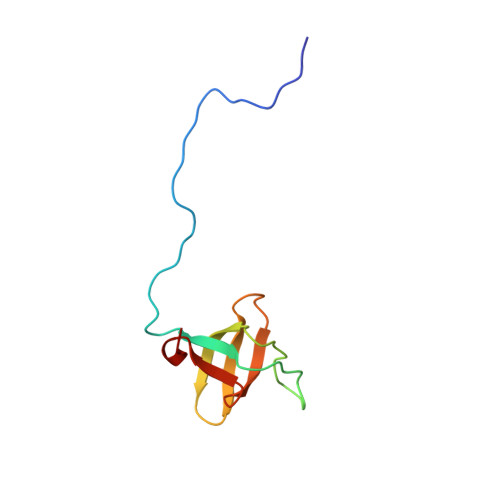
Solution Structure of a Hck SH3 Domain Ligand Complex Reveals Novel Interaction Modes

Schmidt, H.Hoffmann, S.Tran, T.Stoldt, M.Stangler, T.Wiesehan, K.Willbold, D.

(2007) J Mol Biol 365: 1517-1532

  • DOI: https://doi.org/10.1016/j.jmb.2006.11.013
  • Primary Citation of Related Structures:  
    2OI3, 2OJ2

  • PubMed Abstract: 

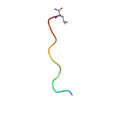
    We studied the interaction of hematopoietic cell kinase SH3 domain (HckSH3) with an artificial 12-residue proline-rich peptide PD1 (HSKYPLPPLPSL) identified as high affinity ligand (K(D)=0.2 muM). PD1 shows an unusual ligand sequence for SH3 binding in type I orientation because it lacks the typical basic anchor residue at position P(-3), but instead has a tyrosine residue at this position. A basic lysine residue, however, is present at position P(-4). The solution structure of the HckSH3:PD1 complex, which is the first HckSH3 complex structure available, clearly reveals that the P(-3) tyrosine residue of PD1 does not take the position of the typical anchor residue but rather forms additional van der Waals interactions with the HckSH3 RT loop. Instead, lysine at position P(-4) of PD1 substitutes the function of the P(-3) anchor residue. This finding expands the well known ligand consensus sequence +xxPpxP by +xxxPpxP. Thus, software tools like iSPOT fail to identify PD1 as a high-affinity HckSH3 ligand so far. In addition, a short antiparallel beta-sheet in the RT loop of HckSH3 is observed upon PD1 binding. The structure of the HckSH3:PD1 complex reveals novel features of SH3 ligand binding and yields new insights into the structural basics of SH3-ligand interactions. Consequences for computational prediction tools adressing SH3-ligand interactions as well as the biological relevance of our findings are discussed.


  • Organizational Affiliation

    Forschungszentrum Jülich GmbH, INB, Biomolecular NMR, 52425 Jülich, Germany.


Macromolecules
Find similar proteins by:  (by identity cutoff)  |  3D Structure
Entity ID: 1
MoleculeChains Sequence LengthOrganismDetailsImage
Tyrosine-protein kinase HCK86Homo sapiensMutation(s): 0 
Gene Names: HCK
EC: 2.7.10.2
UniProt & NIH Common Fund Data Resources
Find proteins for P08631 (Homo sapiens)
Explore P08631 
Go to UniProtKB:  P08631
PHAROS:  P08631
GTEx:  ENSG00000101336 
Entity Groups  
Sequence Clusters30% Identity50% Identity70% Identity90% Identity95% Identity100% Identity
UniProt GroupP08631
Sequence Annotations
Expand
  • Reference Sequence

Find similar proteins by:  Sequence   |   3D Structure  

Entity ID: 2
MoleculeChains Sequence LengthOrganismDetailsImage
artificial peptide PD114N/AMutation(s): 0 
Entity Groups  
Sequence Clusters30% Identity50% Identity70% Identity90% Identity95% Identity100% Identity
Sequence Annotations
Expand
  • Reference Sequence
Experimental Data & Validation

Experimental Data

  • Method: SOLUTION NMR
  • Conformers Calculated: 20 
  • Conformers Submitted: 
  • Selection Criteria: Minimized average structure 

Structure Validation

View Full Validation Report


Entry History 

Deposition Data

Revision History  (Full details and data files)

  • Version 1.0: 2007-02-20
    Type: Initial release
  • Version 1.1: 2008-05-01
    Changes: Version format compliance
  • Version 1.2: 2011-07-13
    Changes: Version format compliance
  • Version 1.3: 2020-02-05
    Changes: Data collection, Database references, Derived calculations, Other
  • Version 1.4: 2023-12-27
    Changes: Data collection, Database references, Derived calculations