Structural Elucidation of the Cell-Penetrating Penetratin Peptide in Model Membranes at the Atomic Level: Probing Hydrophobic Interactions in the Blood-Brain Barrier
Bera, S., Kar, R.K., Mondal, S., Pahan, K., Bhunia, A.(2016) Biochemistry 55: 4982-4996
- PubMed: 27532224
- DOI: https://doi.org/10.1021/acs.biochem.6b00518
- Primary Citation of Related Structures:
2ND6, 2ND7, 2ND8 - PubMed Abstract:
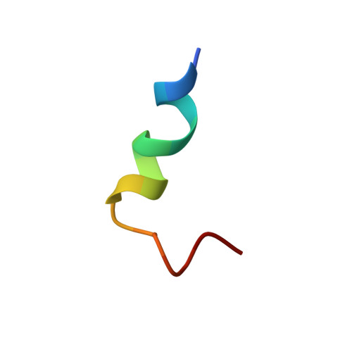
Cell-penetrating peptides (CPPs) have shown promise in nonpermeable therapeutic drug delivery, because of their ability to transport a variety of cargo molecules across the cell membranes and their noncytotoxicity. Drosophila antennapedia homeodomain-derived CPP penetratin (RQIKIWFQNRRMKWKK), being rich in positively charged residues, has been increasingly used as a potential drug carrier for various purposes. Penetratin can breach the tight endothelial network known as the blood-brain barrier (BBB), permitting treatment of several neurodegenerative maladies, including Alzheimer's disease, Parkinson's disease, and Huntington's disease. However, a detailed structural understanding of penetratin and its mechanism of action is lacking. This study defines structural features of the penetratin-derived peptide, DK17 (DRQIKIWFQNRRMKWKK), in several model membranes and describes a membrane-induced conformational transition of the DK17 peptide in these environments. A series of biophysical experiments, including high-resolution nuclear magnetic resonance spectroscopy, provides the three-dimensional structure of DK17 in different membranes mimicking the BBB or total brain lipid extract. Molecular dynamics simulations support the experimental results showing preferential binding of DK17 to particular lipids at atomic resolution. The peptide conserves the structure of the subdomain spanning residues Ile6-Arg11, despite considerable conformational variation in different membrane models. In vivo data suggest that the wild type, not a mutated sequence, enters the central nervous system. Together, these data highlight important structural and functional attributes of DK17 that could be utilized in drug delivery for neurodegenerative disorders.
- Department of Biophysics, Bose Institute , P-1/12 CIT Scheme VII(M), Kolkata 700054, India.
Organizational Affiliation: