From molecular phylogeny towards differentiating pharmacology for NMDA receptor subtypes.
Platt, R.J., Curtice, K.J., Twede, V.D., Watkins, M., Gruszczynski, P., Bulaj, G., Horvath, M.P., Olivera, B.M.(2014) Toxicon 81C: 67-79
- PubMed: 24508768
- DOI: https://doi.org/10.1016/j.toxicon.2014.01.016
- Primary Citation of Related Structures:
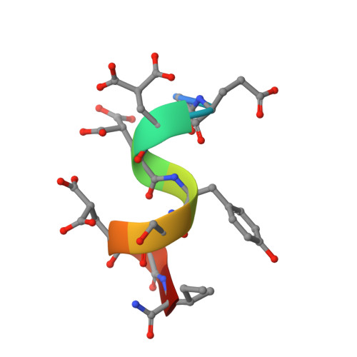
2M7R - PubMed Abstract:
In order to decode the roles that N-methyl-D-aspartate (NMDA) receptors play in excitatory neurotransmission, synaptic plasticity, and neuropathologies, there is need for ligands that differ in their subtype selectivity. The conantokin family of Conus peptides is the only group of peptidic natural products known to target NMDA receptors. Using a search that was guided by phylogeny, we identified new conantokins from the marine snail Conus bocki that complement the current repertoire of NMDA receptor pharmacology. Channel currents measured in Xenopus oocytes demonstrate conantokins conBk-A, conBk-B, and conBk-C have highest potencies for NR2D containing receptors, in contrast to previously characterized conantokins that preferentially block NR2B containing NMDA receptors. Conantokins are rich in γ-carboxyglutamate, typically 17-34 residues, and adopt helical structure in a calcium-dependent manner. As judged by CD spectroscopy, conBk-C adopts significant helical structure in a calcium ion-dependent manner, while calcium, on its own, appears insufficient to stabilize helical conformations of conBk-A or conBk-B. Molecular dynamics simulations help explain the differences in calcium-stabilized structures. Two-dimensional NMR spectroscopy shows that the 9-residue conBk-B is relatively unstructured but forms a helix in the presence of TFE and calcium ions that is similar to other conantokin structures. These newly discovered conantokins hold promise that further exploration of small peptidic antagonists will lead to a set of pharmacological tools that can be used to characterize the role of NMDA receptors in nervous system function and disease.
- Department of Biology, University of Utah, Salt Lake City, UT 84112, USA.
Organizational Affiliation: