Functional Manipulation of a Calcium-binding Protein from Entamoeba histolytica Guided by Paramagnetic NMR.
Rout, A.K., Patel, S., Somlata, Shukla, M., Saraswathi, D., Bhattacharya, A., Chary, K.V.(2013) J Biological Chem 288: 23473-23487
- PubMed: 23782698
- DOI: https://doi.org/10.1074/jbc.M112.411058
- Primary Citation of Related Structures:
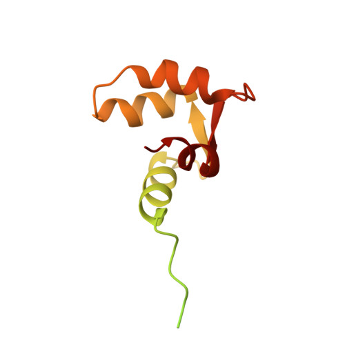
2M7K, 2M7M, 2M7N - PubMed Abstract:
EhCaBP1, one of the calcium-binding proteins from Entamoeba histolytica, is a two-domain EF-hand protein. The two domains of EhCaBP1 are structurally and functionally different from each other. However, both domains are required for structural stability and a full range of functional diversity. Analysis of sequence and structure of EhCaBP1 and other CaBPs indicates that the C-terminal domain of EhCaBP1 possesses a unique structure compared with other family members. This had been attributed to the absence of a Phe-Phe interaction between highly conserved Phe residues at the -4 position in EF-hand III (F[-4]; Tyr(81)) and at the 13th position in EF-hand IV (F[+13]; Phe(129)) of the C-terminal domain. Against this backdrop, we mutated the Tyr residue at the -4th position of EF III to the Phe residue (Y81F), to bring in the Phe-Phe interaction and understand the nature of structural and functional changes in the protein by NMR spectroscopy, molecular dynamics (MD) simulation, isothermal titration calorimetry (ITC), and biological assays, such as imaging and actin binding. The Y81F mutation in EhCaBP1 resulted in a more compact structure for the C-terminal domain of the mutant as in the case of calmodulin and troponin C. The compact structure is favored by the presence of a π-π interaction between Phe(81) and Phe(129) along with several hydrophobic interactions of Phe(81), which are not seen in the wild-type protein. Furthermore, the biological assays reveal preferential membrane localization of the mutant, loss of its colocalization with actin in the phagocytic cups, whereas retaining its ability to bind G- and F-actin.
- Tata Institute of Fundamental Research, Mumbai, India.
Organizational Affiliation: