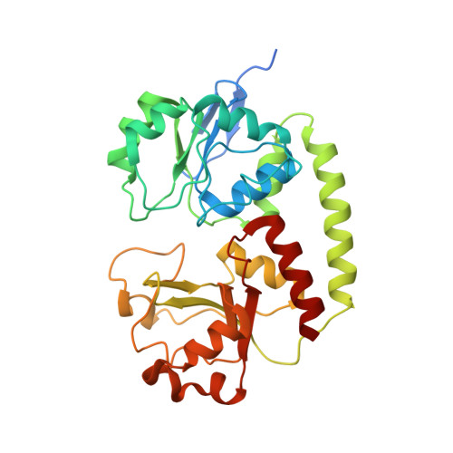
The solution structure, binding properties, and dynamics of the bacterial siderophore-binding protein FepB.
Chu, B.C., Otten, R., Krewulak, K.D., Mulder, F.A., Vogel, H.J.(2014) J Biological Chem 289: 29219-29234
- PubMed: 25173704
- DOI: https://doi.org/10.1074/jbc.M114.564021
- Primary Citation of Related Structures:
2M6K, 2M6L - PubMed Abstract:
The periplasmic binding protein (PBP) FepB plays a key role in transporting the catecholate siderophore ferric enterobactin from the outer to the inner membrane in Gram-negative bacteria. The solution structures of the 34-kDa apo- and holo-FepB from Escherichia coli, solved by NMR, represent the first solution structures determined for the type III class of PBPs. Unlike type I and II PBPs, which undergo large "Venus flytrap" conformational changes upon ligand binding, both forms of FepB maintain similar overall folds; however, binding of the ligand is accompanied by significant loop movements. Reverse methyl cross-saturation experiments corroborated chemical shift perturbation results and uniquely defined the binding pocket for gallium enterobactin (GaEnt). NMR relaxation experiments indicated that a flexible loop (residues 225-250) adopted a more rigid and extended conformation upon ligand binding, which positioned residues for optimal interactions with the ligand and the cytoplasmic membrane ABC transporter (FepCD), respectively. In conclusion, this work highlights the pivotal role that structural dynamics plays in ligand binding and transporter interactions in type III PBPs.
- From the Biochemistry Research Group, Department of Biological Sciences, University of Calgary, Alberta T2N 1N4, Canada.
Organizational Affiliation: