Structure of the L-leucine-binding protein refined at 2.4 A resolution and comparison with the Leu/Ile/Val-binding protein structure.
Sack, J.S., Trakhanov, S.D., Tsigannik, I.H., Quiocho, F.A.(1989) J Mol Biology 206: 193-207
- PubMed: 2649683
- DOI: https://doi.org/10.1016/0022-2836(89)90532-9
- Primary Citation of Related Structures:
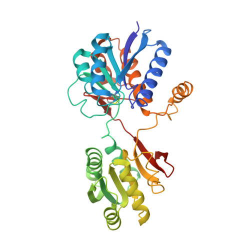
2LBP - PubMed Abstract:
The three-dimensional X-ray structure of the leucine-binding protein (36,900 Mr and 346 residues), an active transport component of Escherichia coli, has been determined by the method of molecular replacement, using the refined structure of the Leu/Ile/Val-binding protein (344 residues) as the model structure. The two amino acid-binding proteins have 80% sequence identity and, although both crystallize in the same space group, they have very different unit cell dimensions. The rotation function yielded one significant peak, which subsequently led to a single self-consistent translation function solution. The model was first refined by the constrained least-squares method, with each of the two domains of the molecule treated separately to allow for any small change in the relative orientation of the two domains. The model was then modified in order to reflect the 72 changes in amino acid side-chains and two insertions in going from the Leu/Ile/Val-binding protein sequence to that of the L-leucine-binding protein. Final structure refinement, using the restrained least-squares technique, resulted in an R-factor of 0.20 for 13,797 reflections to a resolution of 2.4 A. The model is comprised of 2600 protein atoms and 91 solvent molecules. The L-leucine-binding protein structure is, as expected, very similar to the Leu/Ile/Val-binding protein structure; both are in the unliganded conformation with the cleft between the two domains wide open and easily accessible. The superimposing of the structures yields a root-mean-square difference of 0.68 A in the alpha-carbon atoms of the 317 equivalent residues. The five regions of the leucine-binding protein structure that differ by more than 1.6 A from the Leu/Ile/Val-binding protein structure are far from the major portion of the ligand-binding site, which is located in one domain of the bilobate protein. Between the structures, there are three differences in the amino acid side-chains that form the major portion of the substrate-binding sites. These substitutions, by themselves, fail to clearly explain the differences in the specificities for branched aliphatic amino acids.
- Department of Biochemistry, Baylor College of Medicine, Houston, TX 77030.
Organizational Affiliation: