Structural basis for sequence specific DNA binding and protein dimerization of HOXA13.
Zhang, Y., Larsen, C.A., Stadler, H.S., Ames, J.B.(2011) PLoS One 6: e23069-e23069
- PubMed: 21829694
- DOI: https://doi.org/10.1371/journal.pone.0023069
- Primary Citation of Related Structures:
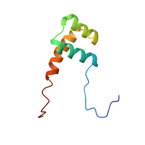
2L7Z, 2LD5 - PubMed Abstract:
The homeobox gene (HOXA13) codes for a transcription factor protein that binds to AT-rich DNA sequences and controls expression of genes during embryonic morphogenesis. Here we present the NMR structure of HOXA13 homeodomain (A13DBD) bound to an 11-mer DNA duplex. A13DBD forms a dimer that binds to DNA with a dissociation constant of 7.5 nM. The A13DBD/DNA complex has a molar mass of 35 kDa consistent with two molecules of DNA bound at both ends of the A13DBD dimer. A13DBD contains an N-terminal arm (residues 324 - 329) that binds in the DNA minor groove, and a C-terminal helix (residues 362 - 382) that contacts the ATAA nucleotide sequence in the major groove. The N370 side-chain forms hydrogen bonds with the purine base of A5* (base paired with T5). Side-chain methyl groups of V373 form hydrophobic contacts with the pyrimidine methyl groups of T5, T6* and T7*, responsible for recognition of TAA in the DNA core. I366 makes similar methyl contacts with T3* and T4*. Mutants (I366A, N370A and V373G) all have decreased DNA binding and transcriptional activity. Exposed protein residues (R337, K343, and F344) make intermolecular contacts at the protein dimer interface. The mutation F344A weakens protein dimerization and lowers transcriptional activity by 76%. We conclude that the non-conserved residue, V373 is critical for structurally recognizing TAA in the major groove, and that HOXA13 dimerization is required to activate transcription of target genes.
- Department of Chemistry, University of California Davis, Davis, California, United States of America.
Organizational Affiliation: