Structure and topology of monomeric phospholamban in lipid membranes determined by a hybrid solution and solid-state NMR approach.
Traaseth, N.J., Shi, L., Verardi, R., Mullen, D.G., Barany, G., Veglia, G.(2009) Proc Natl Acad Sci U S A 106: 10165-10170
- PubMed: 19509339
- DOI: https://doi.org/10.1073/pnas.0904290106
- Primary Citation of Related Structures:
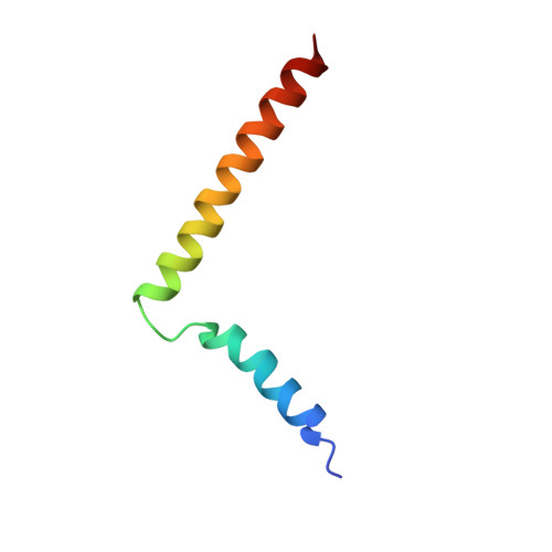
2KB7 - PubMed Abstract:
Phospholamban (PLN) is an essential regulator of cardiac muscle contractility. The homopentameric assembly of PLN is the reservoir for active monomers that, upon deoligomerization form 1:1 complexes with the sarco(endo)plasmic reticulum Ca(2+)-ATPase (SERCA), thus modulating the rate of calcium uptake. In lipid bilayers and micelles, monomeric PLN exists in equilibrium between a bent (or resting) T state and a more dynamic (or active) R state. Here, we report the high-resolution structure and topology of the T state of a monomeric PLN mutant in lipid bilayers, using a hybrid of solution and solid-state NMR restraints together with molecular dynamics simulations in explicit lipid environments. Unlike the previous structural ensemble determined in micelles, this approach gives a complete picture of the PLN monomer structure in a lipid bilayer. This hybrid ensemble exemplifies the tilt, rotation, and depth of membrane insertion, revealing the interaction with the lipids for all protein domains. The N-terminal amphipathic helical domain Ia (residues 1-16) rests on the surface of the lipid membrane with the hydrophobic face of domain Ia embedded in the membrane bilayer interior. The helix comprised of domain Ib (residues 23-30) and transmembrane domain II (residues 31-52) traverses the bilayer with a tilt angle of approximately 24 degrees . The specific interactions between PLN and lipid membranes may represent an additional regulatory element of its inhibitory function. We propose this hybrid method for the simultaneous determination of structure and topology for membrane proteins with compact folds or proteins whose spatial arrangement is dictated by their specific interactions with lipid bilayers.
- Department of Chemistry and Biochemistry, University of Minnesota, Minneapolis, MN 55455, USA.
Organizational Affiliation: