2JA4

Crystal structure of CD5 domain III reveals the fold of a group B scavenger cysteine-rich receptor


Experimental Data Snapshot

  • Method: X-RAY DIFFRACTION
  • Resolution: 2.21 Å
  • R-Value Free: 0.248 
  • R-Value Work: 0.193 
  • R-Value Observed: 0.196 

wwPDB Validation   3D Report Full Report


This is version 1.3 of the entry. See complete history


Literature

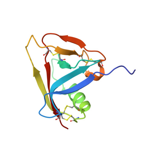
Crystal Structure of the Third Extracellular Domain of Cd5 Reveals the Fold of a Group B Scavenger Cysteine-Rich Receptor Domain.

Rodamilans, B.Munoz, I.G.Bragado-Nilsson, E.Sarrias, M.R.Padilla, O.Blanco, F.J.Lozano, F.Montoya, G.

(2007) J Biol Chem 282: 12669

  • DOI: https://doi.org/10.1074/jbc.M611699200
  • Primary Citation of Related Structures:  
    2JA4, 2OTT

  • PubMed Abstract: 

    Scavenger receptor cysteine-rich (SRCR) domains are ancient protein modules widely found among cell surface and secreted proteins of the innate and adaptive immune system, where they mediate ligand binding. We have solved the crystal structure at 2.2 A of resolution of the SRCR CD5 domain III, a human lymphocyte receptor involved in the modulation of antigen specific receptor-mediated T cell activation and differentiation signals. The first structure of a member of a group B SRCR domain reveals the fold of this ancient protein module into a central core formed by two antiparallel beta-sheets and one alpha-helix, illustrating the conserved core at the protein level of genes coding for group A and B members of the SRCR superfamily. The novel SRCR group B structure permits the interpretation of site-directed mutagenesis data on the binding of activated leukocyte cell adhesion molecule (ALCAM/CD166) binding to CD6, a closely related lymphocyte receptor homologue to CD5.


  • Organizational Affiliation

    Spanish National Cancer Center (CNIO), Structural Biology and Biocomputing Program, c/Melchor Fernandez Almagro 3, 28029 Madrid, Spain.


Macromolecules
Find similar proteins by:  (by identity cutoff)  |  3D Structure
Entity ID: 1
MoleculeChains Sequence LengthOrganismDetailsImage
T-CELL SURFACE GLYCOPROTEIN CD5101Homo sapiensMutation(s): 0 
UniProt & NIH Common Fund Data Resources
Find proteins for P06127 (Homo sapiens)
Explore P06127 
Go to UniProtKB:  P06127
PHAROS:  P06127
GTEx:  ENSG00000110448 
Entity Groups  
Sequence Clusters30% Identity50% Identity70% Identity90% Identity95% Identity100% Identity
UniProt GroupP06127
Sequence Annotations
Expand
  • Reference Sequence
Experimental Data & Validation

Experimental Data

  • Method: X-RAY DIFFRACTION
  • Resolution: 2.21 Å
  • R-Value Free: 0.248 
  • R-Value Work: 0.193 
  • R-Value Observed: 0.196 
  • Space Group: P 32 2 1
Unit Cell:
Length ( Å )Angle ( ˚ )
a = 62.931α = 90
b = 62.931β = 90
c = 72.564γ = 120
Software Package:
Software NamePurpose
ARP/wARPmodel building
SCALEPACKdata scaling
BNPphasing
SHARPphasing
SOLOMONphasing
ARP/wARPphasing
REFMACrefinement

Structure Validation

View Full Validation Report


Entry History 

Deposition Data

Revision History  (Full details and data files)

  • Version 1.0: 2007-03-06
    Type: Initial release
  • Version 1.1: 2011-05-08
    Changes: Version format compliance
  • Version 1.2: 2011-07-13
    Changes: Version format compliance
  • Version 1.3: 2017-06-28
    Changes: Refinement description