2IY4

X-ray structure of Dps from Listeria monocytogenes


Experimental Data Snapshot

  • Method: X-RAY DIFFRACTION
  • Resolution: 2.31 Å
  • R-Value Free: 0.238 
  • R-Value Work: 0.182 
  • R-Value Observed: 0.185 

wwPDB Validation   3D Report Full Report


This is version 1.3 of the entry. See complete history


Literature

The Mutations Lys 114 --> Gln and Asp 126 --> Asn Disrupt an Intersubunit Salt Bridge and Convert Listeria Innocua Dps Into its Natural Mutant Listeria Monocytogenes Dps. Effects on Protein Stability at Low Ph.

Bellapadrona, G.Chiaraluce, R.Consalvi, V.Ilari, A.Stefanini, S.Chiancone, E.

(2007) Proteins 66: 975

  • DOI: https://doi.org/10.1002/prot.21305
  • Primary Citation of Related Structures:  
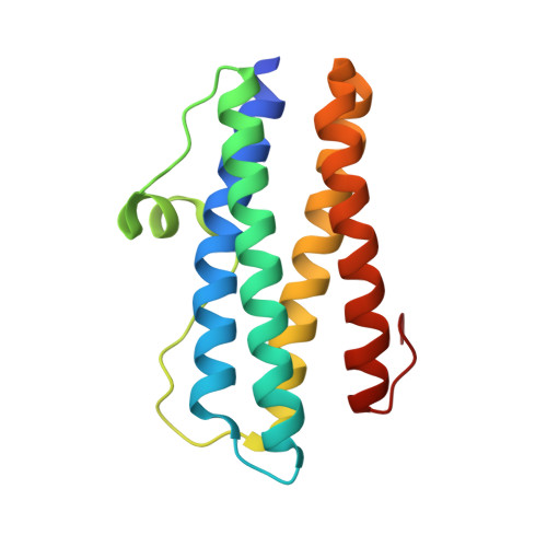
    2IY4

  • PubMed Abstract: 

    The stability of the dodecameric Listeria monocytogenes Dps has been compared with that of the Listeria innocua protein. The two proteins differ only in two amino acid residues that form an intersubunit salt-bridge in L. innocua Dps. This salt-bridge is replaced by a hydrogen bonding network in L. monocytogenes Dps as revealed by the X-ray crystal structure. The resistance to low pH and high temperature was assayed for both Dps proteins under equilibrium conditions and kinetically. Despite the identical equilibrium behavior, significant differences in the kinetic stability and activation energy of the unfolding process are apparent at pH 1.5. The higher stability of L. monocytogenes Dps has been accounted for in terms of the persistence of the hydrogen bonding network at this low pH value. In contrast, the salt-bridge between Lys 114 and Asp 126 characteristic of L. innocua Dps is most likely abolished due to protonation of Asp 126.


  • Organizational Affiliation

    Dipartimento di Scienze Biochimiche A. Rossi Fanelli, Università La Sapienza, Rome, Italy.


Macromolecules
Find similar proteins by:  (by identity cutoff)  |  3D Structure
Entity ID: 1
MoleculeChains Sequence LengthOrganismDetailsImage
NON-HEME IRON-CONTAINING FERRITIN
A,
B,
C,
D,
E,
F,
G,
H,
I,
J,
K,
L,
M,
N,
O,
P,
Q,
R,
S,
T,
U,
V,
W [auth X],
X [auth Y]
156Listeria monocytogenesMutation(s): 0 
UniProt
Find proteins for Q8Y8G1 (Listeria monocytogenes serovar 1/2a (strain ATCC BAA-679 / EGD-e))
Explore Q8Y8G1 
Go to UniProtKB:  Q8Y8G1
Entity Groups  
Sequence Clusters30% Identity50% Identity70% Identity90% Identity95% Identity100% Identity
UniProt GroupQ8Y8G1
Sequence Annotations
Expand
  • Reference Sequence
Small Molecules
Ligands 1 Unique
IDChains Name / Formula / InChI Key2D Diagram3D Interactions
FE
Query on FE

Download Ideal Coordinates CCD File 
AA [auth D]
BA [auth E]
CA [auth E]
DA [auth F]
EA [auth H]
AA [auth D],
BA [auth E],
CA [auth E],
DA [auth F],
EA [auth H],
FA [auth I],
GA [auth J],
HA [auth K],
IA [auth L],
JA [auth M],
KA [auth M],
LA [auth N],
MA [auth N],
NA [auth P],
OA [auth Q],
PA [auth R],
QA [auth R],
RA [auth S],
SA [auth U],
TA [auth V],
UA [auth X],
VA [auth Y],
Y [auth B],
Z [auth C]
FE (III) ION
Fe
VTLYFUHAOXGGBS-UHFFFAOYSA-N
Experimental Data & Validation

Experimental Data

  • Method: X-RAY DIFFRACTION
  • Resolution: 2.31 Å
  • R-Value Free: 0.238 
  • R-Value Work: 0.182 
  • R-Value Observed: 0.185 
  • Space Group: P 1 21 1
Unit Cell:
Length ( Å )Angle ( ˚ )
a = 89.288α = 90
b = 172.708β = 92.11
c = 135.312γ = 90
Software Package:
Software NamePurpose
REFMACrefinement

Structure Validation

View Full Validation Report


Entry History 

Deposition Data

Revision History  (Full details and data files)

  • Version 1.0: 2007-01-02
    Type: Initial release
  • Version 1.1: 2011-05-08
    Changes: Version format compliance
  • Version 1.2: 2011-07-13
    Changes: Version format compliance
  • Version 1.3: 2023-12-13
    Changes: Data collection, Database references, Derived calculations, Other, Refinement description