The solution structure of a domain from the Neisseria meningitidis lipoprotein PilP reveals a new beta-sandwich fold.
Golovanov, A.P., Balasingham, S., Tzitzilonis, C., Goult, B.T., Lian, L.Y., Homberset, H., Tonjum, T., Derrick, J.P.(2006) J Mol Biology 364: 186-195
- PubMed: 17007878
- DOI: https://doi.org/10.1016/j.jmb.2006.08.078
- Primary Citation of Related Structures:
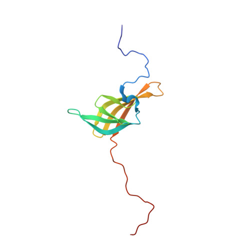
2IVW - PubMed Abstract:
Type IV pili are long, thin fibres, which extend from the surface of the bacterial pathogen Neisseria meningitidis; they play a key role in adhesion and colonisation of host cells. PilP is a lipoprotein, suggested to be involved in the assembly and stabilization of an outer membrane protein, PilQ, which is required for pilus formation. Here we describe the expression of a recombinant fragment of PilP, spanning residues 20 to 181, and determination of the solution structure of a folded domain, spanning residues 85 to 163, by NMR. The N-terminal third of the protein, from residues 20 to 84, is apparently unfolded. Protease digestion yielded a 113 residue fragment that contained the folded domain. The domain adopts a simple beta-sandwich type fold, consisting of a three-stranded beta-sheet packed against a four-stranded beta-sheet. There is also a short segment of 3(10) helix at the N-terminal part of the folded domain. We were unable to identify any other proteins that are closely related in structure to the PilP domain, although the fold appears to be distantly related to the lipocalin family. Over 40 homologues of PilP have been identified in Gram-negative bacteria and the majority of conserved residues lie within the folded domain. The fourth beta-strand and adjacent loop regions contain a high proportion of conserved residues, including three glycine residues, which seem to play a role in linking the two beta-sheets. The two beta-sheets pack together to form a crevice, lined with conserved hydrophobic residues: we suggest that this feature could act as a binding site for a small ligand. The results show that PilP and its homologues have a conserved, folded domain at the C-terminal end of the protein that may be involved in mediating binding to hydrophobic ligands.
- Faculty of Life Sciences and Manchester Interdisciplinary Biocentre, The University of Manchester, 131 Princess Street, Manchester M1 7DN, UK. A.Golovanov@manchester.ac.uk
Organizational Affiliation: