Dynamic docking of cytochrome b5 with myoglobin and alpha-hemoglobin: heme-neutralization "squares" and the binding of electron-transfer-reactive configurations.
Wheeler, K.E., Nocek, J.M., Cull, D.A., Yatsunyk, L.A., Rosenzweig, A.C., Hoffman, B.M.(2007) J Am Chem Soc 129: 3906-3917
- PubMed: 17343378
- DOI: https://doi.org/10.1021/ja067598g
- Primary Citation of Related Structures:
2IN4 - PubMed Abstract:
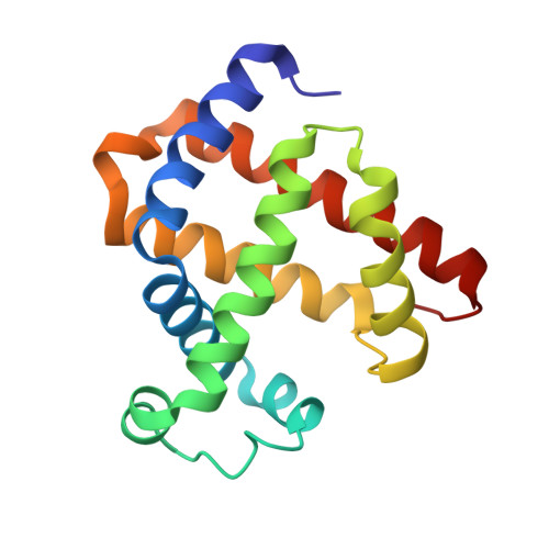
Intracomplex electron transfer (ET) occurs most often in intrinsically transient, low affinity complexes. As a result, the means by which adequate specificity and reactivity are obtained to support effective ET is still poorly understood. We report here on two such ET complexes: cytochrome b5 (cyt b5) in reaction with its physiological partners, myoglobin (Mb) and hemoglobin (Hb). These complexes obey the Dynamic Docking (DD) paradigm: a large ensemble of weakly bound protein-protein configurations contribute to binding in the rapid-exchange limit, but only a few are ET-active. We report the ionic-strength dependence of the second-order rate constant, k2, for photoinitiated ET from within all four combinations of heme-neutralized Zn deuteroporphyrin-substituted Mb/alphaHb undergoing ET with cyt b5, the four "corners" of a "heme-neutralization square". These experiments provide insights into the relative importance of both global and local electrostatic contributions to the binding of reactive configurations, which are too few to be observed directly. To interpret the variations of k2 arising from heme neutralization, we have developed a procedure by which comparisons of the ET rate constants for a heme-neutralization square permit us to decompose the free energy of reactive binding into individual local electrostatic contributions associated with interactions between (i) the propionates of the two hemes and (ii) the heme of each protein with the polypeptide of its partner. Most notably, we find the contribution from the repulsion between propionates of partner hemes to the reactive binding free energy to be surprisingly small, DeltaG(Hb) approximately +1 kcal/mol at ambient temperature, 18 mM ionic strength, and we speculate about possible causes of this observation. To confirm the fundamental assumption of these studies, that the structure of a heme-neutralized protein is unaltered either by substitution of Zn or by heme neutralization, we have obtained the X-ray structure of ZnMb prepared with the porphyrin dimethyl ester and find it to be nearly isostructural with the native protein.
- Department of Chemistry and Department of Biochemistry, Molecular Biology and Cell Biology, Northwestern University, 2145 North Sheridan Road, Evanston, IL 60208, USA.
Organizational Affiliation: