2HTN

E. coli bacterioferritin in its as-isolated form


Experimental Data Snapshot

  • Method: X-RAY DIFFRACTION
  • Resolution: 2.50 Å
  • R-Value Free: 0.188 
  • R-Value Work: 0.171 
  • R-Value Observed: 0.172 

wwPDB Validation   3D Report Full Report


Ligand Structure Quality Assessment 


This is version 1.4 of the entry. See complete history


Literature

Fortuitous structure determination of 'as-isolated' Escherichia coli bacterioferritin in a novel crystal form.

van Eerde, A.Wolterink-van Loo, S.van der Oost, J.Dijkstra, B.W.

(2006) Acta Crystallogr Sect F Struct Biol Cryst Commun 62: 1061-1066

  • DOI: https://doi.org/10.1107/S1744309106039583
  • Primary Citation of Related Structures:  
    2HTN

  • PubMed Abstract: 

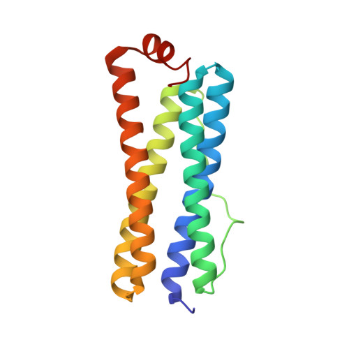
    Escherichia coli bacterioferritin was serendipitously crystallized in a novel cubic crystal form and its structure could be determined to 2.5 A resolution despite a high degree of merohedral twinning. This is the first report of crystallographic data on 'as-isolated' E. coli bacterioferritin. The ferroxidase active site contains positive difference density consistent with two metal ions that had co-purified with the protein. X-ray fluorescence studies suggest that the metal composition is different from that of previous structures and is a mix of zinc and native iron ions. The ferroxidase-centre configuration displays a similar flexibility as previously noted for other bacterioferritins.


  • Organizational Affiliation

    Laboratory of Biophysical Chemistry, University of Groningen, Nijenborgh 4, 9747 AG Groningen, The Netherlands.


Macromolecules
Find similar proteins by:  (by identity cutoff)  |  3D Structure
Entity ID: 1
MoleculeChains Sequence LengthOrganismDetailsImage
Bacterioferritin
A, B, C, D, E
A, B, C, D, E, F, G, H
158Escherichia coli BL21Mutation(s): 0 
EC: 1.16.3.1
UniProt
Find proteins for P0ABD3 (Escherichia coli (strain K12))
Explore P0ABD3 
Go to UniProtKB:  P0ABD3
Entity Groups  
Sequence Clusters30% Identity50% Identity70% Identity90% Identity95% Identity100% Identity
UniProt GroupP0ABD3
Sequence Annotations
Expand
  • Reference Sequence
Small Molecules
Ligands 2 Unique
IDChains Name / Formula / InChI Key2D Diagram3D Interactions
HEM
Query on HEM

Download Ideal Coordinates CCD File 
K [auth A],
P [auth C],
U [auth E],
Z [auth G]
PROTOPORPHYRIN IX CONTAINING FE
C34 H32 Fe N4 O4
KABFMIBPWCXCRK-RGGAHWMASA-L
FE
Query on FE

Download Ideal Coordinates CCD File 
AA [auth H]
BA [auth H]
I [auth A]
J [auth A]
L [auth B]
AA [auth H],
BA [auth H],
I [auth A],
J [auth A],
L [auth B],
M [auth B],
N [auth C],
O [auth C],
Q [auth D],
R [auth D],
S [auth E],
T [auth E],
V [auth F],
W [auth F],
X [auth G],
Y [auth G]
FE (III) ION
Fe
VTLYFUHAOXGGBS-UHFFFAOYSA-N
Experimental Data & Validation

Experimental Data

  • Method: X-RAY DIFFRACTION
  • Resolution: 2.50 Å
  • R-Value Free: 0.188 
  • R-Value Work: 0.171 
  • R-Value Observed: 0.172 
  • Space Group: P 21 3
Unit Cell:
Length ( Å )Angle ( ˚ )
a = 167.893α = 90
b = 167.893β = 90
c = 167.893γ = 90
Software Package:
Software NamePurpose
REFMACrefinement
MOSFLMdata reduction
CCP4data scaling
PHASERphasing

Structure Validation

View Full Validation Report


Ligand Structure Quality Assessment 


Entry History 

Deposition Data

Revision History  (Full details and data files)

  • Version 1.0: 2006-11-21
    Type: Initial release
  • Version 1.1: 2008-05-01
    Changes: Version format compliance
  • Version 1.2: 2011-07-13
    Changes: Derived calculations, Source and taxonomy, Version format compliance
  • Version 1.3: 2014-11-12
    Changes: Derived calculations
  • Version 1.4: 2023-08-30
    Changes: Data collection, Database references, Derived calculations, Refinement description