Crystal structure of oxidized flavodoxin from a red alga Chondrus crispus refined at 1.8 A resolution. Description of the flavin mononucleotide binding site
Fukuyama, K., Matsubara, H., Rogers, L.J.(1992) J Mol Biology 225: 775-789
- PubMed: 1602481
- DOI: https://doi.org/10.1016/0022-2836(92)90400-e
- Primary Citation of Related Structures:
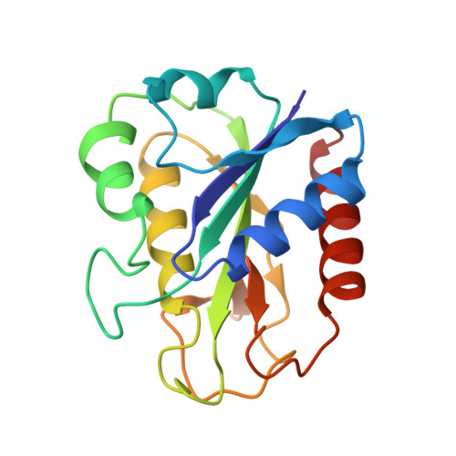
2FCR - PubMed Abstract:
In order to describe the detailed conformation of the oxidized flavodoxin from a eukaryotic red alga, Chondrus crispus, the crystal structure has been refined by a restrained least-squares method. The crystallographic R factor is 0.168 for 13,899 reflections with F greater than 2 sigma F between 6.0 and 1.8 A resolution. The refined model includes 173 amino acid residues, flavin mononucleotide (FMN) and 110 water molecules. The root-mean-square deviation in bond lengths from ideal values is 0.015 A, and the mean co-ordinate error is estimated to be 0.2 A. The FMN is located at the periphery of the molecule. The orientation of the isoalloxazine ring is such that the C-7 and C-8 methyl groups are exposed to solvent and the pyrimidine moiety is buried in the protein. Three peptide segments, T8-T13, T55-T58 and D94-C103, are involved in FMN binding. The first segment of T8-T13 enfolds the phosphate group of the FMN. The three oxygen atoms in the phosphate group form extensive hydrogen bonds with amide groups of the main chain and the O gamma atoms of the side-chains in this segment. T55 O and W56 N epsilon 1 in the second segment form hydrogen bonds with O-2 in the ribityl moiety and one of the oxygen atoms in the phosphate group, respectively. The O gamma H of T58 forms a hydrogen bond with the N-5 atom in the isoalloxazine ring, which is expected to be protonated in the semiquinone form. The third segment is in contact with the isoalloxazine ring. It appears that the hydrogen bond acceptor of the NH of Asp94 in the third segment is O-2 rather than N-1 in the isoalloxazine ring. The isoalloxazine ring is flanked by the side-chains of Trp56 and Tyr98; it forms an angle of 38 degrees with the indole ring of Trp56 and is almost parallel to the benzene ring of Tyr98. The environment of the phosphate group is conserved as in other flavodoxins whereas that of the isoalloxazine ring differs. The relationship between the hydrogen bond to the N-5 in the ring and the redox potential for the oxidized/semiquinone couple is discussed.
- Department of Biology, Faculty of Science, Osaka University, Japan.
Organizational Affiliation: