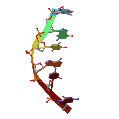
Interactions between morpholinyl anthracyclines and DNA. The crystal structure of a morpholino doxorubicin bound to d(CGTACG).
Cirilli, M., Bachechi, F., Ughetto, G., Colonna, F.P., Capobianco, M.L.(1993) J Mol Biology 230: 878-889
- PubMed: 8478940
- DOI: https://doi.org/10.1006/jmbi.1993.1208
- Primary Citation of Related Structures:
2DES - PubMed Abstract:
Anthracycline antibiotics daunomycin and adriamycin are among the most widely used in cancer chemotherapy and DNA is believed to be the primary target of their biological action. The crystal structure of a morpholino derivative of adriamycin bound to the DNA hexamer d(CGTACG) has been determined at 1.5 A resolution. The complex crystallizes in space group P1 with unit cell dimensions a = 18.01 A, b = 18.83 A, c = 27.65 A, alpha = 92.6 degrees, beta = 100.5 degrees, gamma = 94.9 degrees and there are two drug molecules bound per duplex. Morpholino derivatives differ greatly from their parent compounds in their biological and pharmacological properties. Structural comparison of this complex with the series of previously reported anthracycline-DNA complexes offers an opportunity for studying relationships between structure and function. The anthracycline chromophore intercalates at the CpG step and DNA distortions from a B-type conformation are similar to those observed in the other DNA-anthracycline complexes. Interactions between drug and DNA show no differences at the intercalation site, while in the minor groove they are significantly affected by the presence of the bulky morpholinyl moiety on the anthracycline amino sugar. The binding site involves four base-pairs and the absence of a positive charge on the amino sugar appears to influence the hydration pattern on both grooves. The two halves of the duplex are symmetrically related by a non-crystallographic 2-fold axis but they are not equivalent. In one half, one magnesium cluster bridges both drug and DNA, further stabilizing the complex.
- Istituto di Strutturistica Chimica CNR, Area della Ricerca di Roma, Italy.
Organizational Affiliation: