2ZFO

Structure of the partially unliganded met state of 400 kDa hemoglobin: Insights into ligand-induced structural changes of giant hemoglobins


Experimental Data Snapshot

  • Method: X-RAY DIFFRACTION
  • Resolution: 1.95 Å
  • R-Value Free: 0.202 
  • R-Value Work: 0.169 

wwPDB Validation   3D Report Full Report


Ligand Structure Quality Assessment 


This is version 1.2 of the entry. See complete history


Literature

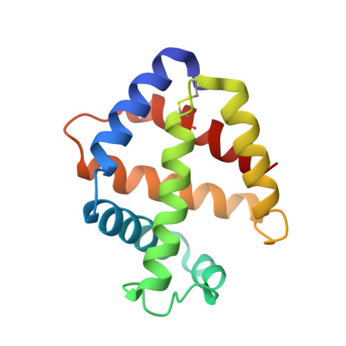
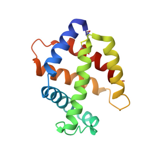
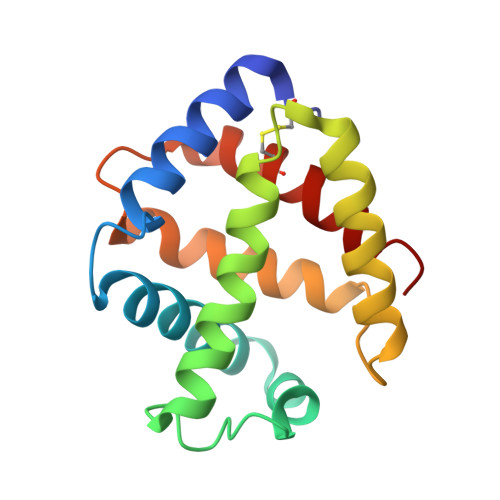
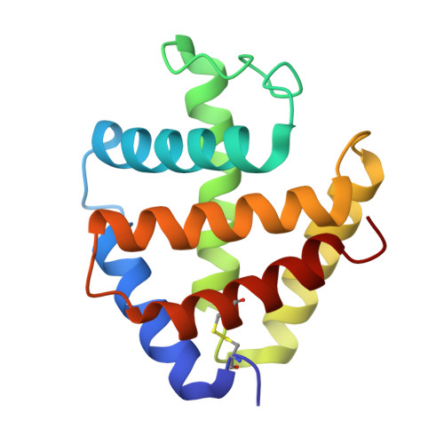
Structure of the partially unliganded met state of 400 kDa hemoglobin: insights into ligand-induced structural changes of giant hemoglobins

Numoto, N.Nakagawa, T.Kita, A.Sasayama, Y.Fukumori, Y.Miki, K.

(2008) Proteins 73: 113-125

  • DOI: https://doi.org/10.1002/prot.22040
  • Primary Citation of Related Structures:  
    2ZFO

  • PubMed Abstract: 

    Recent crystallographic studies have revealed the structures of some invertebrate extracellular giant hemoglobins of 3,600 kDa or 400 kDa and their common quaternary structure of dodecameric subassembly composed of four kinds of globin subunits (A1, A2, B1, and B2). These results have provided insight into the mechanisms of their unique functional properties of oxygen binding and sulfide binding. All of these structures were solved with oxygenated or CO-liganded forms at low or moderate resolutions. We have determined the crystal structure of 400 kDa Hb from a polychaete Oligobrachia mashikoi at 1.95 A resolution. The electron densities at higher resolution confirm the existence of an isoform of the B1 subunit because of the inconsistency with the model that was built from the formerly known amino acid sequence. The brownish color of the crystals used in this study and the absorption spectrum from the dissolved crystals strongly indicated that the obtained structure was a ferric met state, whereas complete absence of electron density around the distal heme pockets were observed at the A2, B1, and B2 subunits. We concluded that the obtained structure was in unliganded met forms at three of four globin subunits in the 24mer assembly and in oxygenated forms at the remaining A1 subunits. The partially unliganded structure showed remarkable structural changes at the AB loop regions causing quaternary rearrangements of the EF-dimer structure. In contrast, few changes occurred at the interface regions composed of the E and F helices. These results suggest that the ligand-induced structural changes of Oligobrachia Hb are quite different from those of the well-studied mollusk Hb having the same EF-dimer structure. The structural rearrangements make the dodecameric subassembly form a tighter conformation than those of fully oxygenated or CO-liganded dodecamer structure.


  • Organizational Affiliation

    Department of Chemistry, Graduate School of Science, Kyoto University, Sakyo-Ku, Kyoto 606-8502, Japan.


Macromolecules
Find similar proteins by:  (by identity cutoff)  |  3D Structure
Entity ID: 1
MoleculeChains Sequence LengthOrganismDetailsImage
Extracellular giant hemoglobin major globin subunit A1140Oligobrachia mashikoiMutation(s): 0 
UniProt
Find proteins for Q7M419 (Oligobrachia mashikoi)
Explore Q7M419 
Go to UniProtKB:  Q7M419
Entity Groups  
Sequence Clusters30% Identity50% Identity70% Identity90% Identity95% Identity100% Identity
UniProt GroupQ7M419
Sequence Annotations
Expand
  • Reference Sequence
Find similar proteins by:  (by identity cutoff)  |  3D Structure
Entity ID: 2
MoleculeChains Sequence LengthOrganismDetailsImage
Extracellular giant hemoglobin major globin subunit A2142Oligobrachia mashikoiMutation(s): 0 
UniProt
Find proteins for Q7M413 (Oligobrachia mashikoi)
Explore Q7M413 
Go to UniProtKB:  Q7M413
Entity Groups  
Sequence Clusters30% Identity50% Identity70% Identity90% Identity95% Identity100% Identity
UniProt GroupQ7M413
Sequence Annotations
Expand
  • Reference Sequence
Find similar proteins by:  (by identity cutoff)  |  3D Structure
Entity ID: 3
MoleculeChains Sequence LengthOrganismDetailsImage
Extracellular giant hemoglobin major globin subunit B2147Oligobrachia mashikoiMutation(s): 0 
UniProt
Find proteins for Q7M418 (Oligobrachia mashikoi)
Explore Q7M418 
Go to UniProtKB:  Q7M418
Entity Groups  
Sequence Clusters30% Identity50% Identity70% Identity90% Identity95% Identity100% Identity
UniProt GroupQ7M418
Sequence Annotations
Expand
  • Reference Sequence
Find similar proteins by:  (by identity cutoff)  |  3D Structure
Entity ID: 4
MoleculeChains Sequence LengthOrganismDetailsImage
Extracellular giant hemoglobin major globin subunit B1145Oligobrachia mashikoiMutation(s): 10 
UniProt
Find proteins for Q5KSB7 (Oligobrachia mashikoi)
Explore Q5KSB7 
Go to UniProtKB:  Q5KSB7
Entity Groups  
Sequence Clusters30% Identity50% Identity70% Identity90% Identity95% Identity100% Identity
UniProt GroupQ5KSB7
Sequence Annotations
Expand
  • Reference Sequence
Experimental Data & Validation

Experimental Data

  • Method: X-RAY DIFFRACTION
  • Resolution: 1.95 Å
  • R-Value Free: 0.202 
  • R-Value Work: 0.169 
  • Space Group: H 3 2
Unit Cell:
Length ( Å )Angle ( ˚ )
a = 110.96α = 90
b = 110.96β = 90
c = 271.58γ = 120
Software Package:
Software NamePurpose
CNSrefinement
BSSdata collection
HKL-2000data reduction
SCALEPACKdata scaling
MOLREPphasing

Structure Validation

View Full Validation Report


Ligand Structure Quality Assessment 


Entry History 

Deposition Data

Revision History  (Full details and data files)

  • Version 1.0: 2008-04-22
    Type: Initial release
  • Version 1.1: 2011-07-13
    Changes: Non-polymer description, Version format compliance
  • Version 1.2: 2023-11-01
    Changes: Data collection, Database references, Derived calculations, Refinement description