Structural Consequences of Nucleophosmin Mutations in Acute Myeloid Leukemia.
Grummitt, C.G., Townsley, F.M., Johnson, C.M., Warren, A.J., Bycroft, M.(2008) J Biological Chem 283: 23326
- PubMed: 18511415
- DOI: https://doi.org/10.1074/jbc.M801706200
- Primary Citation of Related Structures:
2VXD - PubMed Abstract:
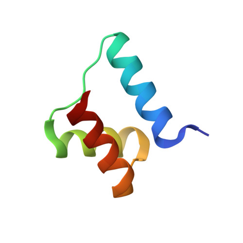
Mutations affecting NPM1 (nucleophosmin) are the most common genetic lesions found in acute myeloid leukemia (AML). NPM1 is one of the most abundant proteins found in the nucleolus and has links to the MDM2/p53 tumor suppressor pathway. A distinctive feature of NPM1 mutants in AML is their aberrant localization to the cytoplasm of leukemic cells. This mutant phenotype is the result of the substitution of several C-terminal residues, including one or two conserved tryptophan residues, with a leucine-rich nuclear export signal. The exact molecular mechanism underlying the loss of nucleolar retention, and the role of the tryptophans, remains unknown. In this study we have determined the structure of an independently folded globular domain in the C terminus of NPM1 using NMR spectroscopy, and we report that the conserved tryptophans are critical for structure. This domain is necessary for the nucleolar targeting of NPM1 and is disrupted by mutations in AML with cytoplasmic NPM1. Furthermore, we identify conserved surface-exposed lysine residues that are functionally rather than structurally important for nucleolar localization. This study provides new focus for efforts to understand the pathogenesis of AML with cytoplasmic NPM1 and may be used to aid the design of small molecules that target the C-terminal domain of NPM1 to act as novel anti-proliferative and anti-leukemia therapeutics.
- Medical Research Council Centre for Protein Engineering, Cambridge CB2 0QH, UK.
Organizational Affiliation: