2V1T

CRYSTAL STRUCTURE OF RAT TOM20-ALDH PRESEQUENCE COMPLEX


Experimental Data Snapshot

  • Method: X-RAY DIFFRACTION
  • Resolution: 1.92 Å
  • R-Value Free: 0.240 
  • R-Value Work: 0.178 
  • R-Value Observed: 0.184 

wwPDB Validation   3D Report Full Report


This is version 1.1 of the entry. See complete history


Literature

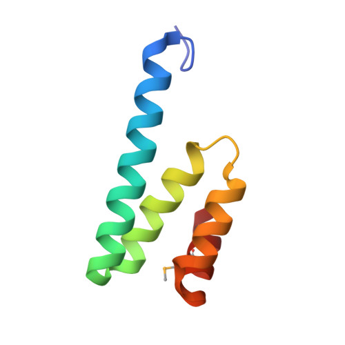
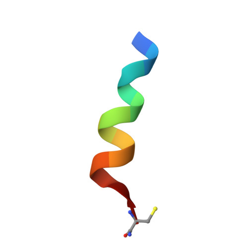
Tom20 Recognizes Mitochondrial Presequences Through Dynamic Equilibrium Among Multiple Bound States.

Saitoh, T.Igura, M.Obita, T.Ose, T.Kojima, R.Maenaka, K.Endo, T.Kohda, D.

(2007) EMBO J 26: 4777

  • DOI: https://doi.org/10.1038/sj.emboj.7601888
  • Primary Citation of Related Structures:  
    2V1S, 2V1T

  • PubMed Abstract: 

    Most mitochondrial proteins are synthesized in the cytosol and imported into mitochondria. The N-terminal presequences of mitochondrial-precursor proteins contain a diverse consensus motif (phi chi chi phi phi, phi is hydrophobic and chi is any amino acid), which is recognized by the Tom20 protein on the mitochondrial surface. To reveal the structural basis of the broad selectivity of Tom20, the Tom20-presequence complex was crystallized. Tethering a presequence peptide to Tom20 through a disulfide bond was essential for crystallization. Unexpectedly, the two crystals with different linker designs provided unique relative orientations of the presequence with respect to Tom20, and neither configuration could fully account for the hydrophobic preference at the three hydrophobic positions of the consensus motif. We propose the existence of a dynamic equilibrium in solution among multiple states including the two bound states. In accordance, NMR 15N relaxation analyses suggested motion on a sub-millisecond timescale at the Tom20-presequence interface. We suggest that the dynamic, multiple-mode interaction is the molecular mechanism facilitating the broadly selective specificity of the Tom20 receptor toward diverse mitochondrial presequences.


  • Organizational Affiliation

    Division of Structural Biology, Medical Institute of Bioregulation, Kyushu University, Maidashi, Higashi-ku, Fukuoka, Japan.


Macromolecules
Find similar proteins by:  (by identity cutoff)  |  3D Structure
Entity ID: 1
MoleculeChains Sequence LengthOrganismDetailsImage
MITOCHONDRIAL IMPORT RECEPTOR SUBUNIT TOM20 HOMOLOG
A, B
73Rattus norvegicusMutation(s): 0 
UniProt
Find proteins for Q62760 (Rattus norvegicus)
Explore Q62760 
Go to UniProtKB:  Q62760
Entity Groups  
Sequence Clusters30% Identity50% Identity70% Identity90% Identity95% Identity100% Identity
UniProt GroupQ62760
Sequence Annotations
Expand
  • Reference Sequence

Find similar proteins by:  Sequence   |   3D Structure  

Entity ID: 2
MoleculeChains Sequence LengthOrganismDetailsImage
ALDEHYDE DEHYDROGENASE
C, D
13Rattus norvegicusMutation(s): 2 
EC: 1.2.1.3
UniProt
Find proteins for P11884 (Rattus norvegicus)
Explore P11884 
Go to UniProtKB:  P11884
Entity Groups  
Sequence Clusters30% Identity50% Identity70% Identity90% Identity95% Identity100% Identity
UniProt GroupP11884
Sequence Annotations
Expand
  • Reference Sequence
Small Molecules
Modified Residues  2 Unique
IDChains TypeFormula2D DiagramParent
MSE
Query on MSE
A, B
L-PEPTIDE LINKINGC5 H11 N O2 SeMET
CY3
Query on CY3
C, D
L-PEPTIDE LINKINGC3 H8 N2 O SCYS
Experimental Data & Validation

Experimental Data

  • Method: X-RAY DIFFRACTION
  • Resolution: 1.92 Å
  • R-Value Free: 0.240 
  • R-Value Work: 0.178 
  • R-Value Observed: 0.184 
  • Space Group: P 1 21 1
Unit Cell:
Length ( Å )Angle ( ˚ )
a = 33.629α = 90
b = 27.639β = 103.07
c = 70.967γ = 90
Software Package:
Software NamePurpose
REFMACrefinement
HKL-2000data reduction
SCALEPACKdata scaling
SOLVEphasing

Structure Validation

View Full Validation Report


Entry History 

Deposition Data

Revision History  (Full details and data files)

  • Version 1.0: 2007-06-12
    Type: Initial release
  • Version 1.1: 2011-07-13
    Changes: Advisory, Version format compliance