2RJW

The crystal structure of the H41Y mutant of villin headpiece, P61 SPACE GROUP.


Experimental Data Snapshot

  • Method: X-RAY DIFFRACTION
  • Resolution: 1.55 Å
  • R-Value Free: 0.229 
  • R-Value Work: 0.202 
  • R-Value Observed: 0.202 

wwPDB Validation   3D Report Full Report


This is version 1.4 of the entry. See complete history


Literature

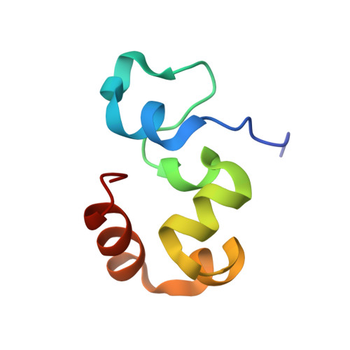
Heterogeneity and dynamics in villin headpiece crystal structures.

Meng, J.McKnight, C.J.

(2009) Acta Crystallogr D Biol Crystallogr 65: 470-476

  • DOI: https://doi.org/10.1107/S0907444909008646
  • Primary Citation of Related Structures:  
    2RJW, 2RJX

  • PubMed Abstract: 

    The villin headpiece domain (HP67) is the C-terminal F-actin-binding motif that confers F-actin-bundling activity to villin, a component of the actin bundles that support the brush-border microvilli. It has been investigated extensively by both experimental and theoretical measurements. Our laboratory, for example, has determined both its NMR and its crystal structures. This study presents the structures of HP67 and its pH-stabilized mutant (H41Y) in a different crystal form and space group. For both constructs, two molecules are found in each asymmetric unit in the new space group P6(1). While one of the two structures (Mol A) is structurally similar to our previously determined structure (Mol X), the other (Mol B) has significant deviations, especially in the N-terminal subdomain, where lattice contacts do not appear to contribute to the difference. In addition, the structurally most different crystal structure, Mol B, is actually closer to the averaged NMR structure. Harmonic motions, as suggested by the B-factor profiles, differ between these crystal structures; crystal structures from the same space group share a similar pattern. Thus, heterogeneity and dynamics are observed in different crystal structures of the same protein even for a protein as small as villin headpiece.


  • Organizational Affiliation

    Boston University School of Medicine, USA.


Macromolecules
Find similar proteins by:  (by identity cutoff)  |  3D Structure
Entity ID: 1
MoleculeChains Sequence LengthOrganismDetailsImage
Villin-1
A, B
67Gallus gallusMutation(s): 1 
Gene Names: VIL1VIL
UniProt
Find proteins for P02640 (Gallus gallus)
Explore P02640 
Go to UniProtKB:  P02640
Entity Groups  
Sequence Clusters30% Identity50% Identity70% Identity90% Identity95% Identity100% Identity
UniProt GroupP02640
Sequence Annotations
Expand
  • Reference Sequence
Experimental Data & Validation

Experimental Data

  • Method: X-RAY DIFFRACTION
  • Resolution: 1.55 Å
  • R-Value Free: 0.229 
  • R-Value Work: 0.202 
  • R-Value Observed: 0.202 
  • Space Group: P 61
Unit Cell:
Length ( Å )Angle ( ˚ )
a = 65.408α = 90
b = 65.408β = 90
c = 67.291γ = 120
Software Package:
Software NamePurpose
CNSrefinement
DENZOdata reduction
SCALEPACKdata scaling
CNSphasing

Structure Validation

View Full Validation Report


Entry History 

Deposition Data

Revision History  (Full details and data files)

  • Version 1.0: 2009-02-17
    Type: Initial release
  • Version 1.1: 2011-07-13
    Changes: Version format compliance
  • Version 1.2: 2021-10-20
    Changes: Database references
  • Version 1.3: 2024-02-21
    Changes: Data collection
  • Version 1.4: 2024-04-03
    Changes: Refinement description