2P8O

Crystal Structure of a Benzohydroxamic Acid/Vanadate complex bound to chymotrypsin A


Experimental Data Snapshot

  • Method: X-RAY DIFFRACTION
  • Resolution: 1.50 Å
  • R-Value Free: 0.243 
  • R-Value Work: 0.199 
  • R-Value Observed: 0.199 

wwPDB Validation   3D Report Full Report


Ligand Structure Quality Assessment 


This is version 1.4 of the entry. See complete history


Literature

Inhibition of chymotrypsin by a complex of ortho-vanadate and benzohydroxamic Acid: structure of the inert complex and its mechanistic interpretation.

Moulin, A.Bell, J.H.Pratt, R.F.Ringe, D.

(2007) Biochemistry 46: 5982-5990

  • DOI: https://doi.org/10.1021/bi6025209
  • Primary Citation of Related Structures:  
    2P8O

  • PubMed Abstract: 

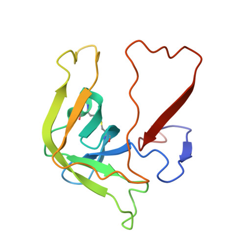
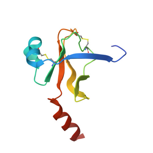
    Serine proteases, like serine beta-lactamases, are rapidly and covalently inhibited by suitably designed phosph(on)ates. The active sites of these enzymes must, therefore, be able to stabilize the pentacoordinated transition states of phosphyl transfer reactions as well as the tetrahedral transition states of acyl transfers. It follows that these enzymes should also be inhibited by molecules capable of generating inert pentacoordinated species. We (J.H.B. and R.F.P.) have previously shown that these enzymes are, in fact, rapidly and reversibly inhibited by 1:1 complexes of vanadate and hydroxamic acids. In this paper, we present the first crystal structure of an acyl transferase inhibited by vanadate. The complex of vanadate and benzohydroxamic acid is a competitive inhibitor of alpha-chymotrypsin with a KI value of 16 muM. In the structure, obtained at a resolution of 1.5 A, the protein is conformationally little different from the apoenzyme. The vanadium, in a distorted octahedral ligand field, is covalently bound to the active site serine oxygen group. One oxgen ligand, presumably anionic, is located in the oxyanion hole. Another is directed roughly in the direction of the acyl transfer leaving group, and a third in the direction of the S2 site. The hydroxamate is bound to vanadium through the hydroxyl oxygen and also, more weakly, through the carbonyl group, to form a five-membered chelate ring. The effect of this chelation is to place the phenyl group of the inhibitor into the important S1 specificity site. The hydroxamate oxygen is directed in line away from the Ser195 Ogamma, approximating the direction of departure of a leaving group in phosphyl transfer. The entire complex can be seen as a reasonable mimic of a phosphyl transfer transition state where the leaving group is extended into the S1 site.


  • Organizational Affiliation

    Rosenstiel Basic Medical Sciences Research Center, Program in Biochemistry, and Program in Biophysics, Brandeis University, Waltham, Massachusetts 02454, USA.


Macromolecules

Find similar proteins by:  Sequence   |   3D Structure  

Entity ID: 1
MoleculeChains Sequence LengthOrganismDetailsImage
Chymotrypsin A chain A13Bos taurusMutation(s): 0 
UniProt
Find proteins for P00766 (Bos taurus)
Explore P00766 
Go to UniProtKB:  P00766
Entity Groups  
Sequence Clusters30% Identity50% Identity70% Identity90% Identity95% Identity100% Identity
UniProt GroupP00766
Sequence Annotations
Expand
  • Reference Sequence
Find similar proteins by:  (by identity cutoff)  |  3D Structure
Entity ID: 2
MoleculeChains Sequence LengthOrganismDetailsImage
Chymotrypsin A chain B131Bos taurusMutation(s): 0 
EC: 3.4.21.1
UniProt
Find proteins for P00766 (Bos taurus)
Explore P00766 
Go to UniProtKB:  P00766
Entity Groups  
Sequence Clusters30% Identity50% Identity70% Identity90% Identity95% Identity100% Identity
UniProt GroupP00766
Sequence Annotations
Expand
  • Reference Sequence
Find similar proteins by:  (by identity cutoff)  |  3D Structure
Entity ID: 3
MoleculeChains Sequence LengthOrganismDetailsImage
Chymotrypsin A chain C97Bos taurusMutation(s): 0 
EC: 3.4.21.1
UniProt
Find proteins for P00766 (Bos taurus)
Explore P00766 
Go to UniProtKB:  P00766
Entity Groups  
Sequence Clusters30% Identity50% Identity70% Identity90% Identity95% Identity100% Identity
UniProt GroupP00766
Sequence Annotations
Expand
  • Reference Sequence
Small Molecules
Ligands 2 Unique
IDChains Name / Formula / InChI Key2D Diagram3D Interactions
BVA
Query on BVA

Download Ideal Coordinates CCD File 
E [auth C]TRIHYDROXY[(N-HYDROXYBENZAMIDATO)OXO]VANADATE
C7 H9 N O5 V
YGVAPMMGCYHYQY-UHFFFAOYSA-K
SO4
Query on SO4

Download Ideal Coordinates CCD File 
D [auth B]SULFATE ION
O4 S
QAOWNCQODCNURD-UHFFFAOYSA-L
Binding Affinity Annotations 
IDSourceBinding Affinity
BVA Binding MOAD:  2P8O Ki: 1.60e+4 (nM) from 1 assay(s)
PDBBind:  2P8O Ki: 1.40e+4 (nM) from 1 assay(s)
Experimental Data & Validation

Experimental Data

  • Method: X-RAY DIFFRACTION
  • Resolution: 1.50 Å
  • R-Value Free: 0.243 
  • R-Value Work: 0.199 
  • R-Value Observed: 0.199 
  • Space Group: P 42 21 2
Unit Cell:
Length ( Å )Angle ( ˚ )
a = 68.989α = 90
b = 68.989β = 90
c = 95.892γ = 90
Software Package:
Software NamePurpose
SHELXrefinement
PDB_EXTRACTdata extraction
HKL-2000data collection
DENZOdata reduction
SCALEPACKdata scaling
CNSphasing
SHELXL-97refinement

Structure Validation

View Full Validation Report


Ligand Structure Quality Assessment 


Entry History 

Deposition Data

Revision History  (Full details and data files)

  • Version 1.0: 2007-05-08
    Type: Initial release
  • Version 1.1: 2008-05-01
    Changes: Version format compliance
  • Version 1.2: 2011-07-13
    Changes: Version format compliance
  • Version 1.3: 2017-10-18
    Changes: Refinement description
  • Version 1.4: 2023-08-30
    Changes: Data collection, Database references, Derived calculations, Refinement description