2OVC

Crystal structure of a coiled-coil tetramerization domain from Kv7.4 channels


Experimental Data Snapshot

  • Method: X-RAY DIFFRACTION
  • Resolution: 2.07 Å
  • R-Value Free: 0.224 
  • R-Value Work: 0.196 
  • R-Value Observed: 0.197 

wwPDB Validation   3D Report Full Report


This is version 1.5 of the entry. See complete history


Literature

Structural Insight into KCNQ (Kv7) Channel Assembly and Channelopathy.

Howard, R.J.Clark, K.A.Holton, J.M.Minor, D.L.

(2007) Neuron 53: 663-675

  • DOI: https://doi.org/10.1016/j.neuron.2007.02.010
  • Primary Citation of Related Structures:  
    2OVC

  • PubMed Abstract: 

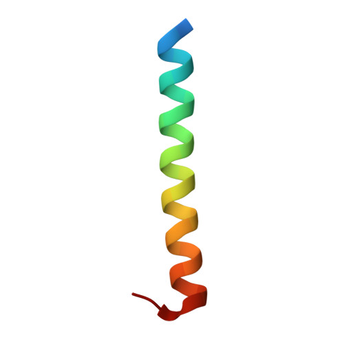
    Kv7.x (KCNQ) voltage-gated potassium channels form the cardiac and auditory I(Ks) current and the neuronal M-current. The five Kv7 subtypes have distinct assembly preferences encoded by a C-terminal cytoplasmic assembly domain, the A-domain Tail. Here, we present the high-resolution structure of the Kv7.4 A-domain Tail together with biochemical experiments that show that the domain is a self-assembling, parallel, four-stranded coiled coil. Structural analysis and biochemical studies indicate conservation of the coiled coil in all Kv7 subtypes and that a limited set of interactions encode assembly specificity determinants. Kv7 mutations have prominent roles in arrhythmias, deafness, and epilepsy. The structure together with biochemical data indicate that A-domain Tail arrhythmia mutations cluster on the solvent-accessible surface of the subunit interface at a likely site of action for modulatory proteins. Together, the data provide a framework for understanding Kv7 assembly specificity and the molecular basis of a distinct set of Kv7 channelopathies.


  • Organizational Affiliation

    Chemistry and Chemical Biology Graduate Program, University of California, San Francisco, CA 94158-2330, USA.


Macromolecules
Find similar proteins by:  (by identity cutoff)  |  3D Structure
Entity ID: 1
MoleculeChains Sequence LengthOrganismDetailsImage
Potassium voltage-gated channel subfamily KQT member 433Homo sapiensMutation(s): 0 
Gene Names: KCNQ4
UniProt & NIH Common Fund Data Resources
Find proteins for P56696 (Homo sapiens)
Explore P56696 
Go to UniProtKB:  P56696
PHAROS:  P56696
GTEx:  ENSG00000117013 
Entity Groups  
Sequence Clusters30% Identity50% Identity70% Identity90% Identity95% Identity100% Identity
UniProt GroupP56696
Sequence Annotations
Expand
  • Reference Sequence
Experimental Data & Validation

Experimental Data

  • Method: X-RAY DIFFRACTION
  • Resolution: 2.07 Å
  • R-Value Free: 0.224 
  • R-Value Work: 0.196 
  • R-Value Observed: 0.197 
  • Space Group: I 4
Unit Cell:
Length ( Å )Angle ( ˚ )
a = 33.185α = 90
b = 33.185β = 90
c = 55.283γ = 90
Software Package:
Software NamePurpose
REFMACrefinement
SCALEPACKdata scaling
CNSrefinement
DENZOdata reduction
PDB_EXTRACTdata extraction
ADSCdata collection
HKL-2000data reduction
HKL-2000data scaling
CNSphasing

Structure Validation

View Full Validation Report


Entry History 

Deposition Data

Revision History  (Full details and data files)

  • Version 1.0: 2007-03-13
    Type: Initial release
  • Version 1.1: 2008-05-01
    Changes: Version format compliance
  • Version 1.2: 2011-07-13
    Changes: Derived calculations, Version format compliance
  • Version 1.3: 2017-10-18
    Changes: Refinement description
  • Version 1.4: 2019-07-24
    Changes: Data collection, Refinement description
  • Version 1.5: 2024-02-21
    Changes: Data collection, Database references