2OHH

Crystal Structure of coenzyme F420H2 oxidase (FprA), a diiron flavoprotein, active oxidized state


Experimental Data Snapshot

  • Method: X-RAY DIFFRACTION
  • Resolution: 1.70 Å
  • R-Value Free: 0.218 
  • R-Value Work: 0.184 
  • R-Value Observed: 0.186 

wwPDB Validation   3D Report Full Report


Ligand Structure Quality Assessment 


This is version 1.3 of the entry. See complete history


Literature

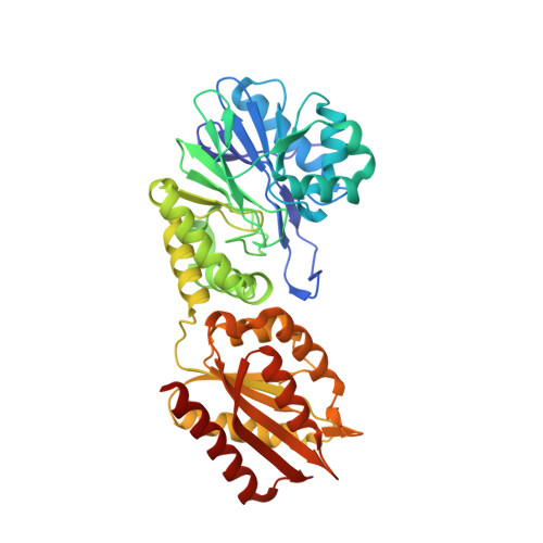
Structure of coenzyme F420H2 oxidase (FprA), a di-iron flavoprotein from methanogenic Archaea catalyzing the reduction of O2 to H2O.

Seedorf, H.Hagemeier, C.H.Shima, S.Thauer, R.K.Warkentin, E.Ermler, U.

(2007) FEBS J 274: 1588-1599

  • DOI: https://doi.org/10.1111/j.1742-4658.2007.05706.x
  • Primary Citation of Related Structures:  
    2OHH, 2OHI, 2OHJ

  • PubMed Abstract: 

    The di-iron flavoprotein F(420)H(2) oxidase found in methanogenic Archaea catalyzes the four-electron reduction of O(2) to 2H(2)O with 2 mol of reduced coenzyme F(420)(7,8-dimethyl-8-hydroxy-5-deazariboflavin). We report here on crystal structures of the homotetrameric F(420)H(2) oxidase from Methanothermobacter marburgensis at resolutions of 2.25 A, 2.25 A and 1.7 A, respectively, from which an active reduced state, an inactive oxidized state and an active oxidized state could be extracted. As found in structurally related A-type flavoproteins, the active site is formed at the dimer interface, where the di-iron center of one monomer is juxtaposed to FMN of the other. In the active reduced state [Fe(II)Fe(II)FMNH(2)], the two irons are surrounded by four histidines, one aspartate, one glutamate and one bridging aspartate. The so-called switch loop is in a closed conformation, thus preventing F(420) binding. In the inactive oxidized state [Fe(III)FMN], the iron nearest to FMN has moved to two remote binding sites, and the switch loop is changed to an open conformation. In the active oxidized state [Fe(III)Fe(III)FMN], both irons are positioned as in the reduced state but the switch loop is found in the open conformation as in the inactive oxidized state. It is proposed that the redox-dependent conformational change of the switch loop ensures alternate complete four-electron O(2) reduction and redox center re-reduction. On the basis of the known Si-Si stereospecific hydride transfer, F(420)H(2) was modeled into the solvent-accessible pocket in front of FMN. The inactive oxidized state might provide the molecular basis for enzyme inactivation by long-term O(2) exposure observed in some members of the FprA family.


  • Organizational Affiliation

    Max Planck Institute for Terrestrial Microbiology, Marburg, Germany.


Macromolecules
Find similar proteins by:  (by identity cutoff)  |  3D Structure
Entity ID: 1
MoleculeChains Sequence LengthOrganismDetailsImage
Type A flavoprotein fprAA,
B,
C [auth D],
D [auth E]
404Methanothermobacter thermautotrophicusMutation(s): 0 
Gene Names: fprAfpaA
EC: 1
UniProt
Find proteins for Q50497 (Methanothermobacter marburgensis (strain ATCC BAA-927 / DSM 2133 / JCM 14651 / NBRC 100331 / OCM 82 / Marburg))
Explore Q50497 
Go to UniProtKB:  Q50497
Entity Groups  
Sequence Clusters30% Identity50% Identity70% Identity90% Identity95% Identity100% Identity
UniProt GroupQ50497
Sequence Annotations
Expand
  • Reference Sequence
Small Molecules
Ligands 3 Unique
IDChains Name / Formula / InChI Key2D Diagram3D Interactions
FMN
Query on FMN

Download Ideal Coordinates CCD File 
G [auth A],
J [auth B],
N [auth D],
Q [auth E]
FLAVIN MONONUCLEOTIDE
C17 H21 N4 O9 P
FVTCRASFADXXNN-SCRDCRAPSA-N
SO4
Query on SO4

Download Ideal Coordinates CCD File 
M [auth D]SULFATE ION
O4 S
QAOWNCQODCNURD-UHFFFAOYSA-L
FE
Query on FE

Download Ideal Coordinates CCD File 
E [auth A]
F [auth A]
H [auth B]
I [auth B]
K [auth D]
E [auth A],
F [auth A],
H [auth B],
I [auth B],
K [auth D],
L [auth D],
O [auth E],
P [auth E]
FE (III) ION
Fe
VTLYFUHAOXGGBS-UHFFFAOYSA-N
Experimental Data & Validation

Experimental Data

  • Method: X-RAY DIFFRACTION
  • Resolution: 1.70 Å
  • R-Value Free: 0.218 
  • R-Value Work: 0.184 
  • R-Value Observed: 0.186 
  • Space Group: P 1 21 1
Unit Cell:
Length ( Å )Angle ( ˚ )
a = 73.74α = 90
b = 120.86β = 110.4
c = 92.69γ = 90
Software Package:
Software NamePurpose
REFMACrefinement
HKL-2000data collection
XDSdata reduction
XDSdata scaling
EPMRphasing

Structure Validation

View Full Validation Report


Ligand Structure Quality Assessment 


Entry History 

Deposition Data

Revision History  (Full details and data files)

  • Version 1.0: 2007-05-22
    Type: Initial release
  • Version 1.1: 2008-05-01
    Changes: Version format compliance
  • Version 1.2: 2011-07-13
    Changes: Advisory, Version format compliance
  • Version 1.3: 2023-12-27
    Changes: Data collection, Database references, Derived calculations