2LBV

Siderocalin Q83 reveals a dual ligand binding mode


Experimental Data Snapshot

  • Method: SOLUTION NMR
  • Conformers Calculated: 200 
  • Conformers Submitted: 20 
  • Selection Criteria: structures with the least restraint violations 

wwPDB Validation   3D Report Full Report


This is version 1.1 of the entry. See complete history


Literature

Lipocalin Q83 reveals a dual ligand binding mode with potential implications for the functions of siderocalins

Coudevylle, N.Hoetzinger, M.Geist, L.Kontaxis, G.Hartl, M.Bister, K.Konrat, R.

(2011) Biochemistry 50: 9192-9199

  • DOI: https://doi.org/10.1021/bi201115q
  • Primary Citation of Related Structures:  
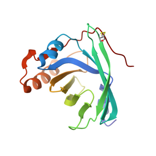
    2LBV

  • PubMed Abstract: 

    Siderocalins are particular lipocalins that participate in the innate immune response by interfering with bacterial siderophore-mediated iron uptake. Additionally, siderocalins are involved in several physiological and pathological processes such as inflammation, iron delivery, tissue differentiation, and cancer progression. Here we show that siderocalin Q83 displays an unexpected dual ligand binding mode as it can bind enterobactin and unsaturated fatty acids simultaneously. The solution structure of the siderocalin Q83 in complex with arachidonic acid and enterobactin reveals molecular details of this novel dual binding mode and the determinants of fatty acid binding specificity. Our results suggest that Q83 is a metabolic hub linking iron and fatty acid pathways. This unexpected coupling might contribute to the pleiotropic functions of siderocalins.


  • Organizational Affiliation

    Department of Structural and Computational Biology, Max F Perutz Laboratories, University of Vienna, Campus Vienna Biocenter 5/1, 1030 Vienna, Austria. nicolas.coudevylle@univie.ac.at


Macromolecules
Find similar proteins by:  (by identity cutoff)  |  3D Structure
Entity ID: 1
MoleculeChains Sequence LengthOrganismDetailsImage
Extracellular fatty acid-binding protein157Coturnix japonicaMutation(s): 0 
UniProt
Find proteins for Q9I9P7 (Coturnix japonica)
Explore Q9I9P7 
Go to UniProtKB:  Q9I9P7
Entity Groups  
Sequence Clusters30% Identity50% Identity70% Identity90% Identity95% Identity100% Identity
UniProt GroupQ9I9P7
Sequence Annotations
Expand
  • Reference Sequence
Small Molecules
Ligands 3 Unique
IDChains Name / Formula / InChI Key2D Diagram3D Interactions
EB4
Query on EB4

Download Ideal Coordinates CCD File 
B [auth A]N,N',N''-[(3S,7S,11S)-2,6,10-trioxo-1,5,9-trioxacyclododecane-3,7,11-triyl]tris(2,3-dihydroxybenzamide)
C30 H27 N3 O15
SERBHKJMVBATSJ-BZSNNMDCSA-N
ACD
Query on ACD

Download Ideal Coordinates CCD File 
D [auth A]ARACHIDONIC ACID
C20 H32 O2
YZXBAPSDXZZRGB-DOFZRALJSA-N
GA
Query on GA

Download Ideal Coordinates CCD File 
C [auth A]GALLIUM (III) ION
Ga
CKHJYUSOUQDYEN-UHFFFAOYSA-N
Binding Affinity Annotations 
IDSourceBinding Affinity
ACD PDBBind:  2LBV Kd: 26 (nM) from 1 assay(s)
Experimental Data & Validation

Experimental Data

  • Method: SOLUTION NMR
  • Conformers Calculated: 200 
  • Conformers Submitted: 20 
  • Selection Criteria: structures with the least restraint violations 

Structure Validation

View Full Validation Report


Entry History 

Deposition Data

Revision History  (Full details and data files)

  • Version 1.0: 2012-02-22
    Type: Initial release
  • Version 1.1: 2023-06-14
    Changes: Database references, Derived calculations, Other