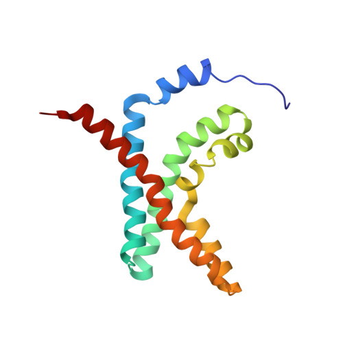
Solution Structure and Phospholipid Interactions of the Isolated Voltage-Sensor Domain from KvAP.
Butterwick, J.A., Mackinnon, R.(2010) J Mol Biology 403: 591-606
- PubMed: 20851706
- DOI: https://doi.org/10.1016/j.jmb.2010.09.012
- Primary Citation of Related Structures:
2KYH - PubMed Abstract:
Voltage-sensor domains (VSDs) are specialized transmembrane segments that confer voltage sensitivity to many proteins such as ion channels and enzymes. The activities of these domains are highly dependent on both the chemical properties and the physical properties of the surrounding membrane environment. To learn about VSD-lipid interactions, we used nuclear magnetic resonance spectroscopy to determine the structure and phospholipid interface of the VSD from the voltage-dependent K(+) channel KvAP (prokaryotic Kv from Aeropyrum pernix). The solution structure of the KvAP VSD solubilized within phospholipid micelles is similar to a previously determined crystal structure solubilized by a nonionic detergent and complexed with an antibody fragment. The differences observed include a previously unidentified short amphipathic α-helix that precedes the first transmembrane helix and a subtle rigid-body repositioning of the S3-S4 voltage-sensor paddle. Using (15)N relaxation experiments, we show that much of the VSD, including the pronounced kink in S3 and the S3-S4 paddle, is relatively rigid on the picosecond-to-nanosecond timescale. In contrast, the kink in S3 is mobile on the microsecond-to-millisecond timescale and may act as a hinge in the movement of the paddle during channel gating. We characterized the VSD-phospholipid micelle interactions using nuclear Overhauser effect spectroscopy and showed that the micelle uniformly coats the KvAP VSD and approximates the chemical environment of a phospholipid bilayer. Using paramagnetically labeled phospholipids, we show that bilayer-forming lipids interact with the S3 and S4 helices more strongly than with S1 and S2.
- Laboratory of Molecular Neurobiology and Biophysics, The Rockefeller University, Howard Hughes Medical Institute, 1230 York Avenue, New York, NY 10065, USA. jbutterwic@rockefeller.edu
Organizational Affiliation: