2KFX

Structure of the N-terminal domain of human cardiac troponin C bound to calcium ion and to the inhibitor W7


Experimental Data Snapshot

  • Method: SOLUTION NMR
  • Conformers Calculated: 50 
  • Conformers Submitted: 10 
  • Selection Criteria: structures with the lowest energy 

wwPDB Validation   3D Report Full Report


This is version 1.2 of the entry. See complete history


Literature

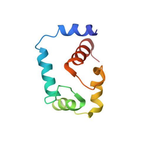
Structure of the inhibitor W7 bound to the regulatory domain of cardiac troponin C.

Hoffman, R.M.Sykes, B.D.

(2009) Biochemistry 48: 5541-5552

  • DOI: https://doi.org/10.1021/bi9001826
  • Primary Citation of Related Structures:  
    2KFX

  • PubMed Abstract: 

    The calmodulin antagonist W7 binds to troponin C in the presence of Ca(2+) and inhibits striated muscle contraction. This study integrates multiple data into the structure of the regulatory domain of human cardiac troponin C (cNTnC) bound to Ca(2+) and W7. The protein-W7 interface is defined through a three-dimensional {(1)H,(13)C}-edited-{(1)H,(12)C}-detected NOESY NMR experiment, and other aspects of the structure are modeled as perturbations to previously known coordinates and restraints. The structure determination protocol optimizes the protein-W7 contacts prior to the introduction of protein-W7 steric interactions or conformational changes in the protein. The structure determination protocol gives families of conformers that all have an optimal docking as assessed by satisfaction of the target function. The structure supports the previously proposed troponin I blocking mechanism for the activity of W7 in striated muscle and suggests a role for the flexible tail of W7 in stabilization of the bound state. This clarifies the structure-activity relationships of W7 and implicates an electrostatically mediated component of activity in common analogues of W7, including the antipsychotic trifluoroperazine and the cardiotonic levosimendan.


  • Organizational Affiliation

    Department of Biochemistry, University of Alberta, Edmonton, Alberta, Canada T6G 2H7.


Macromolecules
Find similar proteins by:  (by identity cutoff)  |  3D Structure
Entity ID: 1
MoleculeChains Sequence LengthOrganismDetailsImage
Troponin C, slow skeletal and cardiac musclesA [auth T]89Homo sapiensMutation(s): 2 
Gene Names: TNNC1TNNC
UniProt & NIH Common Fund Data Resources
Find proteins for P63316 (Homo sapiens)
Explore P63316 
Go to UniProtKB:  P63316
PHAROS:  P63316
GTEx:  ENSG00000114854 
Entity Groups  
Sequence Clusters30% Identity50% Identity70% Identity90% Identity95% Identity100% Identity
UniProt GroupP63316
Sequence Annotations
Expand
  • Reference Sequence
Small Molecules
Ligands 2 Unique
IDChains Name / Formula / InChI Key2D Diagram3D Interactions
WW7
Query on WW7

Download Ideal Coordinates CCD File 
C [auth T]N-(6-AMINOHEXYL)-5-CHLORO-1-NAPHTHALENESULFONAMIDE
C16 H21 Cl N2 O2 S
IDEHCMNLNCJQST-UHFFFAOYSA-N
CA
Query on CA

Download Ideal Coordinates CCD File 
B [auth T]CALCIUM ION
Ca
BHPQYMZQTOCNFJ-UHFFFAOYSA-N
Binding Affinity Annotations 
IDSourceBinding Affinity
WW7 PDBBind:  2KFX Kd: 1.00e+5 (nM) from 1 assay(s)
Experimental Data & Validation

Experimental Data

  • Method: SOLUTION NMR
  • Conformers Calculated: 50 
  • Conformers Submitted: 10 
  • Selection Criteria: structures with the lowest energy 

Structure Validation

View Full Validation Report


Entry History 

Deposition Data

Revision History  (Full details and data files)

  • Version 1.0: 2009-06-09
    Type: Initial release
  • Version 1.1: 2011-07-13
    Changes: Version format compliance
  • Version 1.2: 2021-10-20
    Changes: Data collection, Database references, Derived calculations