Pressure-dependent structure changes in barnase on ligand binding reveal intermediate rate fluctuations.
Wilton, D.J., Kitahara, R., Akasaka, K., Pandya, M.J., Williamson, M.P.(2009) Biophys J 97: 1482-1490
- PubMed: 19720037
- DOI: https://doi.org/10.1016/j.bpj.2009.06.022
- Primary Citation of Related Structures:
2KF3, 2KF4, 2KF5, 2KF6 - PubMed Abstract:
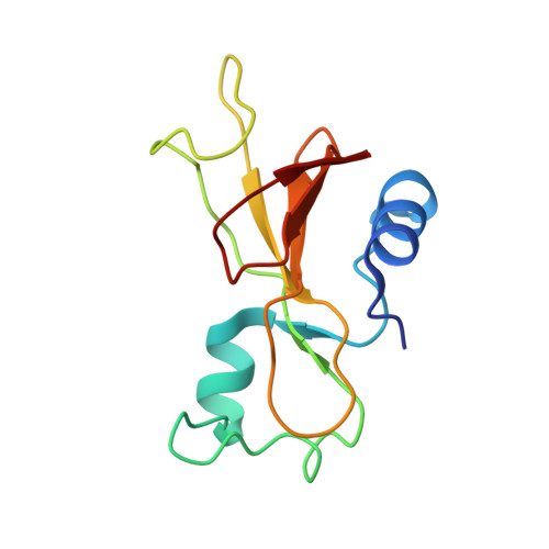
In this work we measured 1H NMR chemical shifts for the ribonuclease barnase at pressures from 3 MPa to 200 MPa, both free and bound to d(CGAC). Shift changes with pressure were used as restraints to determine the change in structure with pressure. Free barnase is compressed by approximately 0.7%. The largest changes are on the ligand-binding face close to Lys-27, which is the recognition site for the cleaved phosphate bond. This part of the protein also contains the buried water molecules. In the presence of d(CGAC), the compressibility is reduced by approximately 70% and the region of structural change is altered: the ligand-binding face is now almost incompressible, whereas changes occur at the opposite face. Because compressibility is proportional to mean square volume fluctuation, we conclude that in free barnase, volume fluctuation is largest close to the active site, but when the inhibitor is bound, the fluctuations become much smaller and are located mainly on the opposite face. The timescale of the fluctuations is nanoseconds to microseconds, consistent with the degree of ordering required for the fluctuations, which are intermediate between rapid uncorrelated side-chain dynamics and slow conformational transitions. The high-pressure technique is therefore useful for characterizing motions on this relatively inaccessible timescale.
- Department of Molecular Biology and Biotechnology, University of Sheffield, Sheffield, United Kingdom.
Organizational Affiliation: