2C6Y

Crystal structure of interleukin enhancer-binding factor 1 bound to DNA


Experimental Data Snapshot

  • Method: X-RAY DIFFRACTION
  • Resolution: 2.40 Å
  • R-Value Free: 0.258 
  • R-Value Work: 0.233 
  • R-Value Observed: 0.233 

wwPDB Validation   3D Report Full Report


This is version 1.2 of the entry. See complete history


Literature

Crystal Structure of the Human Foxk1A-DNA Complex and its Implications on the Diverse Binding Specificity of Winged Helix/Forkhead Proteins.

Tsai, K.-L.Huang, C.-Y.Chang, C.-H.Sun, Y.-J.Chuang, W.-J.Hsiao, C.-D.

(2006) J Biol Chem 281: 17400

  • DOI: https://doi.org/10.1074/jbc.M600478200
  • Primary Citation of Related Structures:  
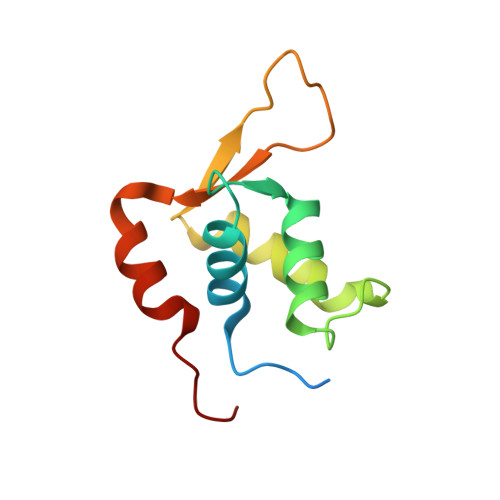
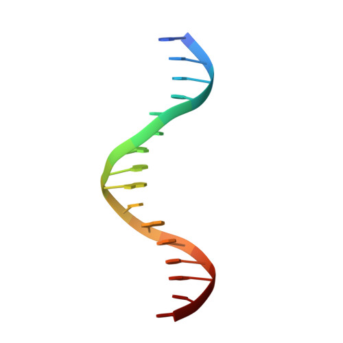
    2C6Y

  • PubMed Abstract: 

    Interleukin enhancer binding factor (ILF) is a human transcription factor and a new member of the winged helix/forkhead family. ILF can bind to purine-rich regulatory motifs such as the human T-cell leukemia virus-long terminal region and the interleukin-2 promoter. Here we report the 2.4 A crystal structure of two DNA binding domains of ILF (FOXK1a) binding to a 16-bp DNA duplex containing a promoter sequence. Electrophoretic mobility shift assay studies demonstrate that two ILF-DNA binding domain molecules cooperatively bind to DNA. In addition to the recognition helix recognizing the core sequences through the major groove, the structure shows that wing 1 interacts with the minor groove of DNA, and the H2-H3 loop region makes ionic bonds to the phosphate group, which permits the recognition of DNA. The structure also reveals that the presence of the C-terminal alpha-helix in place of a typical wing 2 in a member of this family alters the orientation of the C-terminal basic residues (RKRRPR) when binding to DNA outside the core sequence. These results provide a new insight into how the DNA binding specificities of winged helix/forkhead proteins may be regulated by their less conserved regions.


  • Organizational Affiliation

    Institute of Molecular Biology, Academia Sinica, Taipei 115; Institute of Bioinformatics and Structural Biology, National Tsing Hua University, Hsinchu 300.


Macromolecules

Find similar proteins by:  (by identity cutoff)  |  3D Structure
Entity ID: 1
MoleculeChains Sequence LengthOrganismDetailsImage
FORKHEAD BOX PROTEIN K2
A, B
111Homo sapiensMutation(s): 0 
UniProt & NIH Common Fund Data Resources
Find proteins for Q01167 (Homo sapiens)
Explore Q01167 
Go to UniProtKB:  Q01167
PHAROS:  Q01167
GTEx:  ENSG00000141568 
Entity Groups  
Sequence Clusters30% Identity50% Identity70% Identity90% Identity95% Identity100% Identity
UniProt GroupQ01167
Sequence Annotations
Expand
  • Reference Sequence

Find similar nucleic acids by:  Sequence   |   3D Structure  

Entity ID: 2
MoleculeChains LengthOrganismImage
INTERLEUKIN 2 PROMOTOR16Homo sapiens
Sequence Annotations
Expand
  • Reference Sequence

Find similar nucleic acids by:  Sequence   |   3D Structure  

Entity ID: 3
MoleculeChains LengthOrganismImage
INTERLEUKIN 2 PROMOTOR16Homo sapiens
Sequence Annotations
Expand
  • Reference Sequence
Experimental Data & Validation

Experimental Data

  • Method: X-RAY DIFFRACTION
  • Resolution: 2.40 Å
  • R-Value Free: 0.258 
  • R-Value Work: 0.233 
  • R-Value Observed: 0.233 
  • Space Group: P 61 2 2
Unit Cell:
Length ( Å )Angle ( ˚ )
a = 58.736α = 90
b = 58.736β = 90
c = 324.923γ = 120
Software Package:
Software NamePurpose
CNSrefinement
HKL-2000data reduction
HKL-2000data scaling
SOLVEphasing

Structure Validation

View Full Validation Report


Entry History 

Deposition Data

Revision History  (Full details and data files)

  • Version 1.0: 2006-04-18
    Type: Initial release
  • Version 1.1: 2011-05-08
    Changes: Version format compliance
  • Version 1.2: 2011-07-13
    Changes: Version format compliance