2A0L

Crystal structure of KvAP-33H1 Fv complex


Experimental Data Snapshot

  • Method: X-RAY DIFFRACTION
  • Resolution: 3.90 Å
  • R-Value Free: 0.392 
  • R-Value Work: 0.358 
  • R-Value Observed: 0.358 

wwPDB Validation   3D Report Full Report


This is version 1.2 of the entry. See complete history


Literature

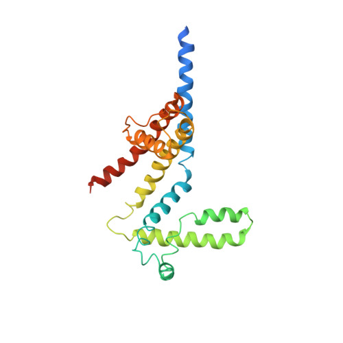
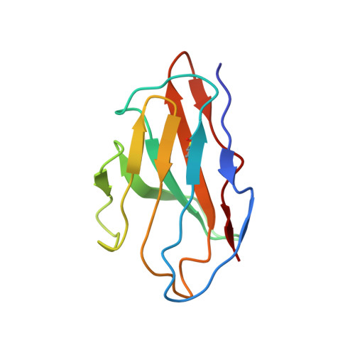
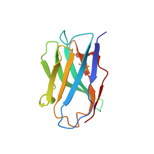
Structure of the KvAP voltage-dependent K+ channel and its dependence on the lipid membrane.

Lee, S.Y.Lee, A.Chen, J.Mackinnon, R.

(2005) Proc Natl Acad Sci U S A 102: 15441-15446

  • DOI: https://doi.org/10.1073/pnas.0507651102
  • Primary Citation of Related Structures:  
    2A0L

  • PubMed Abstract: 

    Voltage-dependent ion channels gate open in response to changes in cell membrane voltage. This form of gating permits the propagation of action potentials. We present two structures of the voltage-dependent K(+) channel KvAP, in complex with monoclonal Fv fragments (3.9 A) and without antibody fragments (8 A). We also studied KvAP with disulfide cross-bridges in lipid membranes. Analyzing these data in the context of the crystal structure of Kv1.2 and EPR data on KvAP we reach the following conclusions: (i) KvAP is similar in structure to Kv1.2 with a very modest difference in the orientation of its voltage sensor; (ii) mAb fragments are not the source of non-native conformations of KvAP in crystal structures; (iii) because KvAP contains separate loosely adherent domains, a lipid membrane is required to maintain their correct relative orientations, and (iv) the model of KvAP is consistent with the proposal of voltage sensing through the movement of an arginine-containing helix-turn-helix element at the protein-lipid interface.


  • Organizational Affiliation

    Howard Hughes Medical Institute, Laboratory of Molecular Neurobiology and Biophysics, The Rockefeller University, 1230 York Avenue, New York, NY 10021, USA.


Macromolecules
Find similar proteins by:  (by identity cutoff)  |  3D Structure
Entity ID: 1
MoleculeChains Sequence LengthOrganismDetailsImage
Voltage-gated potassium channel
A, B
241Aeropyrum pernixMutation(s): 0 
Gene Names: KVAP_AERPE
Membrane Entity: Yes 
UniProt
Find proteins for Q9YDF8 (Aeropyrum pernix (strain ATCC 700893 / DSM 11879 / JCM 9820 / NBRC 100138 / K1))
Explore Q9YDF8 
Go to UniProtKB:  Q9YDF8
Entity Groups  
Sequence Clusters30% Identity50% Identity70% Identity90% Identity95% Identity100% Identity
UniProt GroupQ9YDF8
Sequence Annotations
Expand
  • Reference Sequence
Find similar proteins by:  (by identity cutoff)  |  3D Structure
Entity ID: 2
MoleculeChains Sequence LengthOrganismDetailsImage
33H1 Fv fragment
C, E
105Mus musculusMutation(s): 0 
Entity Groups  
Sequence Clusters30% Identity50% Identity70% Identity90% Identity95% Identity100% Identity
Sequence Annotations
Expand
  • Reference Sequence
Find similar proteins by:  (by identity cutoff)  |  3D Structure
Entity ID: 3
MoleculeChains Sequence LengthOrganismDetailsImage
33H1 Fv fragment
D, F
116Mus musculusMutation(s): 0 
Entity Groups  
Sequence Clusters30% Identity50% Identity70% Identity90% Identity95% Identity100% Identity
Sequence Annotations
Expand
  • Reference Sequence
Experimental Data & Validation

Experimental Data

  • Method: X-RAY DIFFRACTION
  • Resolution: 3.90 Å
  • R-Value Free: 0.392 
  • R-Value Work: 0.358 
  • R-Value Observed: 0.358 
  • Space Group: P 42 2 2
Unit Cell:
Length ( Å )Angle ( ˚ )
a = 136.362α = 90
b = 136.362β = 90
c = 191.812γ = 90
Software Package:
Software NamePurpose
CNSrefinement
MOSFLMdata reduction
CCP4data scaling
MLPHAREphasing

Structure Validation

View Full Validation Report


Entry History 

Deposition Data

Revision History  (Full details and data files)

  • Version 1.0: 2005-11-01
    Type: Initial release
  • Version 1.1: 2008-04-30
    Changes: Version format compliance
  • Version 1.2: 2011-07-13
    Changes: Version format compliance