28MB | pdb_000028mb

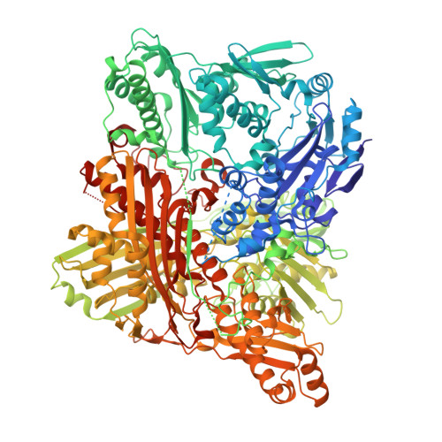
Structure of Human Aldehyde oxidase under TCEP-reducing conditions


Experimental Data Snapshot

  • Method: X-RAY DIFFRACTION
  • Resolution: 2.31 Å
  • R-Value Free: 
    0.241 (Depositor), 0.241 (DCC) 
  • R-Value Work: 
    0.209 (Depositor), 0.210 (DCC) 

Starting Model: experimental
View more details

wwPDB Validation 3D Report Full Report

Validation slider image for 28MB

Ligand Structure Quality Assessment 


This is version 1.3 of the entry. See complete history

Literature

Structure of human aldehyde oxidase under tris(2-carboxyethyl)phosphine reducing conditions.

Videira, C.Esmaeeli, M.Leimkuhler, S.Romao, M.J.Mota, C.

(2026) Acta Crystallogr F Struct Biol Commun 82: 275-282

  • DOI: https://doi.org/10.1107/S2053230X26006904
  • Primary Citation Related Structures: 
    28MB

  • PubMed Abstract: 

    The importance of human aldehyde oxidase (hAOX1) has increased in recent decades due to its involvement in drug metabolism. Inhibition studies involving hAOX1 are extensive and a common reducing agent, dithiothreitol (DTT), was recently found to inactivate the enzyme. However, in previous crystallographic studies of hAOX1, DTT was found to be essential for crystallization. To surpass this concern, another reducing agent was used in crystallization trials. Using tris(2-carboxyethyl)phosphine (TCEP), a sulfur-free reducing agent, it was possible to obtain well ordered crystals of wild-type hAOX1 and a variant, hAOX1_6A, which diffracted beyond 2.3 Å resolution. Instead of the typical star-shaped crystals of hAOX1, at pH 4.7 plates are obtained in the orthorhombic space group P2 1 2 1 2 with two molecules in the asymmetric unit. Activity assays with the enzyme incubated with both reducing agents show that in contrast to DTT, TCEP did not inactivate hAOX1. The replacement of DTT with TCEP in the crystallization of hAOX1 provides a strategy to circumvent enzyme inactivation during crystallographic studies, allowing future applications of new assays, such as time-resolved crystallography.


  • Organizational Affiliation
    • UCIBIO, Applied Molecular Biosciences Unit and Associate Laboratory i4HB - Institute for Health and Bioeconomy, Department of Chemistry NOVA School of Science and Technology, Universidade Nova de Lisboa, 2829-516 Caparica, Portugal.

Macromolecule Content 

  • Total Structure Weight: 299.7 kDa 
  • Atom Count: 20,291 
  • Modeled Residue Count: 2,583 
  • Deposited Residue Count: 2,676 
  • Unique protein chains: 1

Macromolecules

Find similar proteins by:|  3D Structure
Entity ID: 1
MoleculeChains  Sequence LengthOrganismDetailsImage
Aldehyde oxidase
A, B
1,338Homo sapiensMutation(s): 6 
Gene Names: AOX1AO
EC: 1.2.3.1 (PDB Primary Data), 1.17.3 (PDB Primary Data)
UniProt & NIH Common Fund Data Resources
Find proteins for Q06278 (Homo sapiens)
Explore Q06278 
Go to UniProtKB:  Q06278
PHAROS:  Q06278
GTEx:  ENSG00000138356 
Entity Groups
Sequence Clusters30% Identity50% Identity70% Identity90% Identity95% Identity100% Identity
UniProt GroupQ06278
Sequence Annotations
Expand
Reference Sequence

Small Molecules

Ligands 6 Unique
IDChains Name / Formula / InChI Key2D Diagram3D Interactions
FAD
(Subject of Investigation/LOI)

Query on FAD


Download:Ideal Coordinates CCD File
E [auth A],
M [auth B]
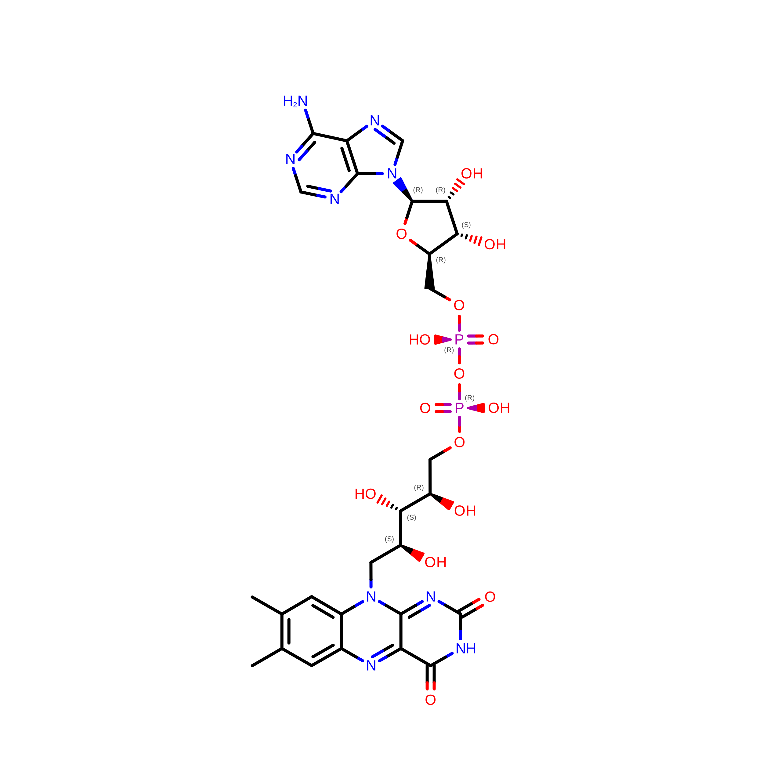
FLAVIN-ADENINE DINUCLEOTIDE
C27 H33 N9 O15 P2
VWWQXMAJTJZDQX-UYBVJOGSSA-N
MTE
(Subject of Investigation/LOI)

Query on MTE


Download:Ideal Coordinates CCD File
I [auth A],
P [auth B]
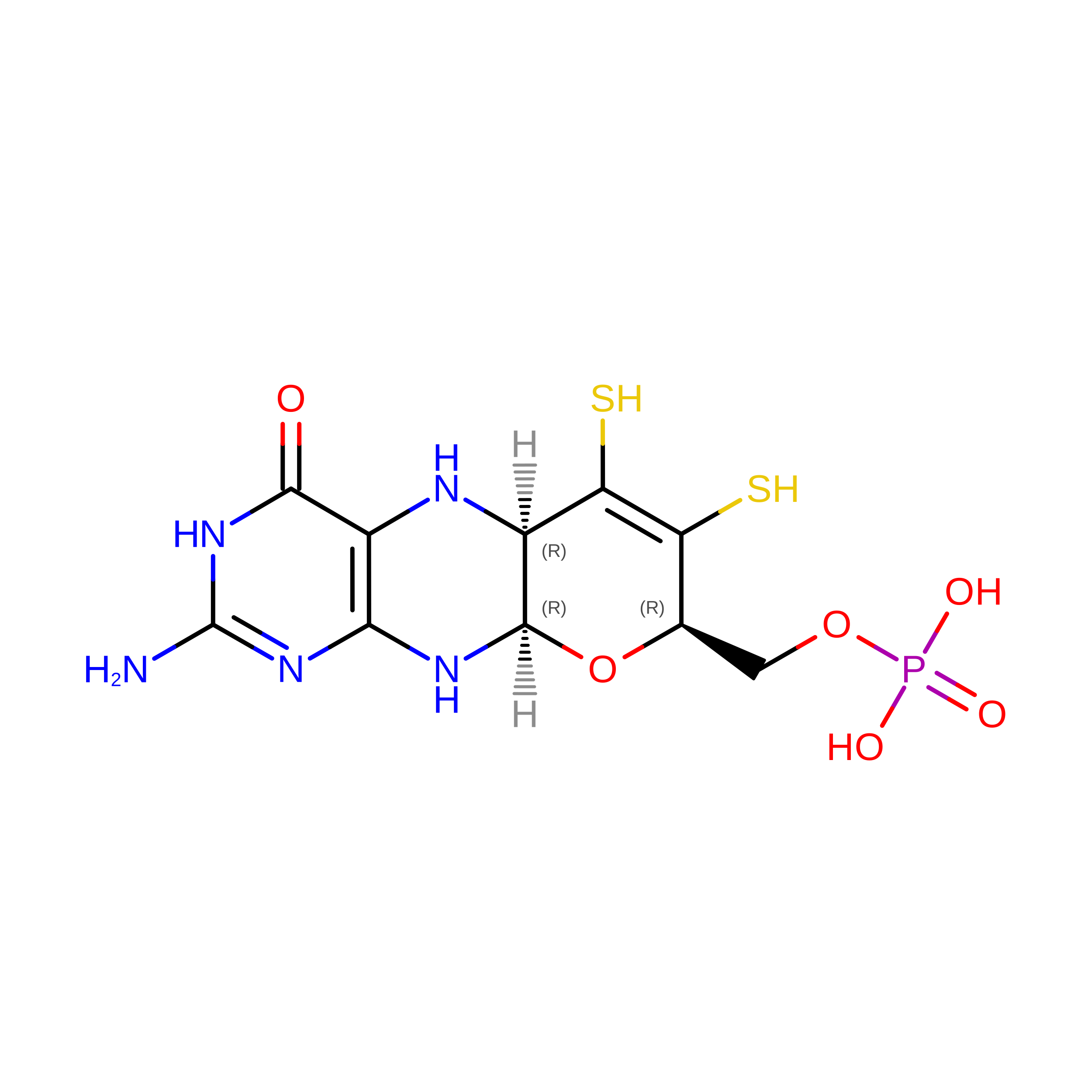
PHOSPHONIC ACIDMONO-(2-AMINO-5,6-DIMERCAPTO-4-OXO-3,7,8A,9,10,10A-HEXAHYDRO-4H-8-OXA-1,3,9,10-TETRAAZA-ANTHRACEN-7-YLMETHYL)ESTER
C10 H14 N5 O6 P S2
HPEUEJRPDGMIMY-IFQPEPLCSA-N
FES
(Subject of Investigation/LOI)

Query on FES


Download:Ideal Coordinates CCD File
C [auth A],
D [auth A],
K [auth B],
L [auth B]
FE2/S2 (INORGANIC) CLUSTER
Fe2 S2
NIXDOXVAJZFRNF-UHFFFAOYSA-N
MOS
(Subject of Investigation/LOI)

Query on MOS


Download:Ideal Coordinates CCD File
J [auth A],
Q [auth B]
DIOXOTHIOMOLYBDENUM(VI) ION
H Mo O2 S
BDSRWPHSAKXXRG-UHFFFAOYSA-M
MLI

Query on MLI


Download:Ideal Coordinates CCD File
F [auth A],
G [auth A],
H [auth A],
O [auth B]
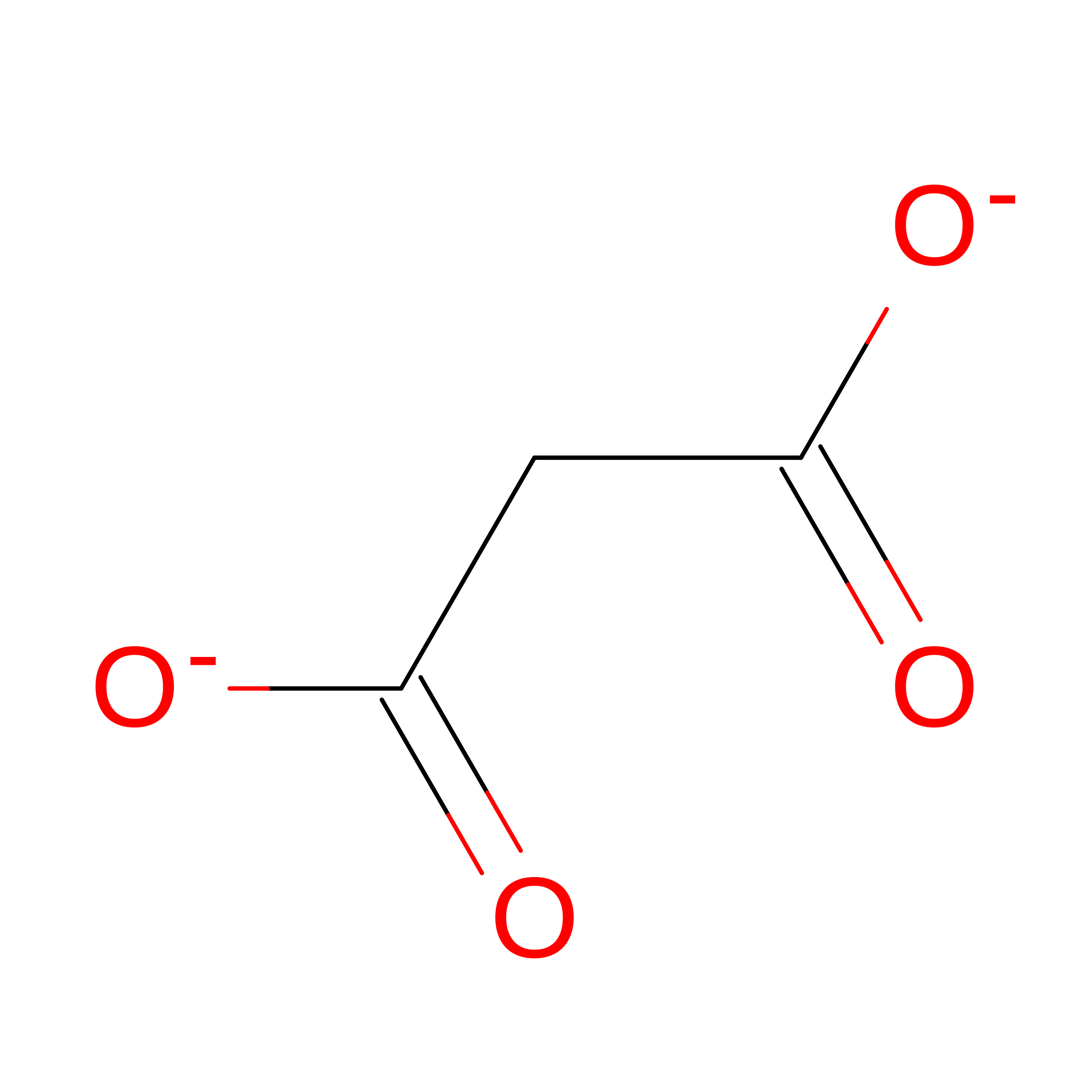
MALONATE ION
C3 H2 O4
OFOBLEOULBTSOW-UHFFFAOYSA-L
GOL

Query on GOL


Download:Ideal Coordinates CCD File
N [auth B]GLYCEROL
C3 H8 O3
PEDCQBHIVMGVHV-UHFFFAOYSA-N

Experimental Data & Validation

Experimental Data

  • Method: X-RAY DIFFRACTION
  • Resolution: 2.31 Å
  • R-Value Free:  0.241 (Depositor), 0.241 (DCC) 
  • R-Value Work:  0.209 (Depositor), 0.210 (DCC) 
Space Group: P 21 21 2
Unit Cell:
Length ( Å )Angle ( ˚ )
a = 191.204α = 90
b = 273.118β = 90
c = 77.883γ = 90
Software Package:
Software NamePurpose
REFMACrefinement
PHASERphasing
autoPROCdata processing
STARANISOdata scaling
XDSdata reduction
Aimlessdata scaling
XSCALEdata scaling
pointlessdata scaling

Structure Validation

View Full Validation Report


Ligand Structure Quality Assessment 


Entry History 

& Funding Information

Deposition Data


Funding OrganizationLocationGrant Number
Fundacao para a Ciencia e a TecnologiaPortugal2023.18077.ICDT
Fundacao para a Ciencia e a TecnologiaPortugalUIDP/04378/2020
Fundacao para a Ciencia e a TecnologiaPortugalUIDB/04378/2020
Fundacao para a Ciencia e a TecnologiaPortugalLA/P/0140/2020

Revision History  (Full details and data files)

  • Version 1.0: 2026-07-29
    Type: Initial release
  • Version 1.1: 2026-08-05
    Changes: Database references
  • Version 1.2: 2026-08-12
    Type: Remediation
    Reason: Metalloprotein remediation
    Changes: Derived calculations, Structure summary
  • Version 1.3: 2026-08-19
    Changes: Database references