






 Download Mendeley
Cryo-EM structure of oat amyloid fibrils reveals ordered histidine arrays on the surface
Li, S., Li, S., Cao, Q., Fang, Y., Cao, Y.(2027) Food Hydrocoll 184: 113342
Experimental Data Snapshot
wwPDB Validation 3D Report Full Report

Entity ID: 1 | |||||
|---|---|---|---|---|---|
| Molecule | Chains | Sequence Length | Organism | Details | Image |
| 11S globulin | 45 | Avena sativa | Mutation(s): 0 Gene Names: GLAV1 | 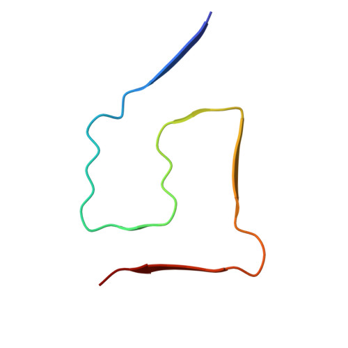 | |
UniProt | |||||
Entity Groups | |||||
| Sequence Clusters | 30% Identity50% Identity70% Identity90% Identity95% Identity100% Identity | ||||
| UniProt Group | Q38779 | ||||
Sequence AnnotationsExpand | |||||
Reference Sequence | |||||
| Task | Software Package | Version |
|---|---|---|
| MODEL REFINEMENT | PHENIX | |
| RECONSTRUCTION | RELION | 4.0 |
| Funding Organization | Location | Grant Number |
|---|---|---|
| Not funded | -- |








