23SS | pdb_000023ss

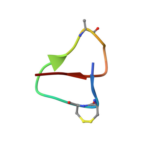
The lasso structure of jiangxilassin A


Experimental Data Snapshot

  • Method: SOLUTION NMR
  • Conformers Calculated: 100 
  • Conformers Submitted: 20 
  • Selection Criteria: target function 

wwPDB Validation 3D Report Full Report

Validation slider image for 23SS

This is version 1.1 of the entry. See complete history. 

Literature

Structural and biochemical analyses of a lasso peptide natural product containing an unusual disulfide bond between consecutive cysteines.

Kawakami, A., Tsunoda, T., Kumeta, H., Maruyama, C., Hamano, Y., Dairi, T., Ogasawara, Y.

(2026) Biosci Biotechnol Biochem 

  • DOI: https://doi.org/10.1093/bbb/zbag135
  • Primary Citation Related Structures: 
    23SS

  • PubMed Abstract: 

    Lasso peptides are a group of ribosomally synthesized and post-translationally modified peptides with a unique lariat topology. They have been classified into four classes based on disulfide bond patterns. In this study, we isolated a lasso peptide containing a disulfide bond in the macrocyclic ring, which is identical to jiangxilassin A. Analysis of the three-dimensional solution structure of this peptide suggested that the disulfide bond enhances the rigidity of the macrocyclic ring. To investigate whether the disulfide bond is required for enzymatic modification, we replaced either cysteine residue with serine. Upon heterologous expression, both mutants produced disulfide-linked dimers that retained macrolactam ring formation, dehydration, and methylation, although their production levels were substantially lower than the level observed for the wild type. These results indicate that the vicinal disulfide bond is not strictly required for these post-translational modifications but contributes to the efficient production of jiangxilassin A.


  • Organizational Affiliation: 
    • Graduate School of Chemical Sciences and Engineering, Hokkaido University, Sapporo, Japan.

Macromolecule Content 

  • Total Structure Weight: 2.01 kDa 
  • Atom Count: 139 
  • Modeled Residue Count: 17 
  • Deposited Residue Count: 17 
  • Unique protein chains: 1

Macromolecules

Find similar proteins by:  Sequence   |   3D Structure  
Entity ID: 1
MoleculeChains  Sequence LengthOrganismDetailsImage
MET-THR-CYS-CYS-GLY-LYS-TRP-ALA-ASP-DHA-ILE-TRP-GLY-TYR-PHE-PHE-ASP17Nonomuraea jiangxiensisMutation(s): 0 
Entity Groups
Sequence Clusters30% Identity50% Identity70% Identity90% Identity95% Identity100% Identity
Sequence Annotations
Expand
Reference Sequence

Small Molecules

Modified Residues  1 Unique
IDChains Modified ResidueModification TypeFormula2D Diagram
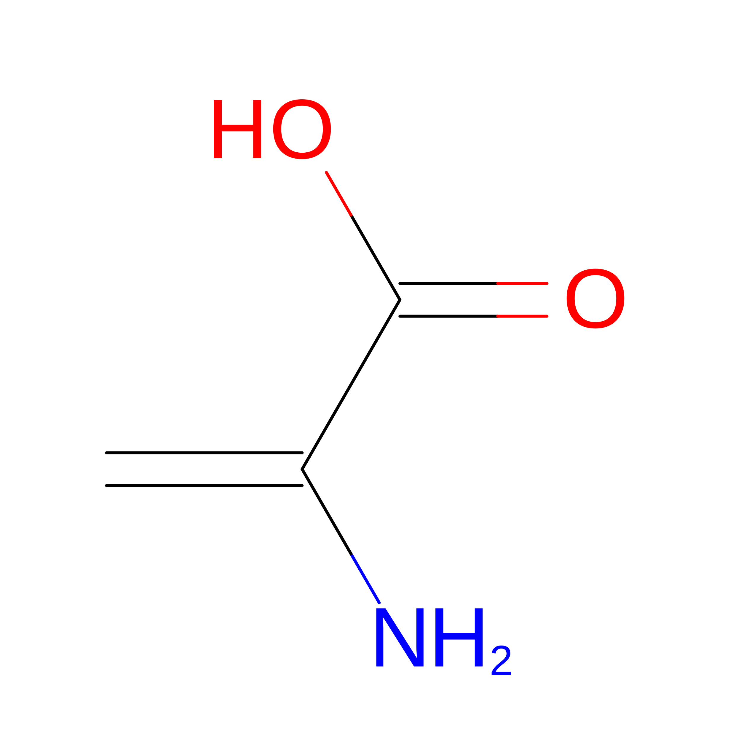
DHA
Query on DHA
SERDehydroamino acidC3 H5 N O2

Experimental Data & Validation

Experimental Data

  • Method: SOLUTION NMR
  • Conformers Calculated: 100 
  • Conformers Submitted: 20 
  • Selection Criteria: target function 

Structure Validation

View Full Validation Report


Entry History 

& Funding Information

Deposition Data


Funding OrganizationLocationGrant Number
Japan Society for the Promotion of Science (JSPS)JapanJP16H06452
Japan Society for the Promotion of Science (JSPS)JapanJP18H03937
Japan Society for the Promotion of Science (JSPS)JapanJP22H04976
Japan Society for the Promotion of Science (JSPS)JapanJP18K05449
Japan Society for the Promotion of Science (JSPS)JapanJP22H05130
Japan Society for the Promotion of Science (JSPS)JapanJP23H02145
Japan Society for the Promotion of Science (JSPS)JapanJP23K19276
Japan Society for the Promotion of Science (JSPS)JapanJP25KJ0506

Revision History  (Full details and data files)

  • Version 1.0: 2026-09-16
    Type: Initial release
  • Version 1.1: 2026-09-23
    Changes: Database references