20XK | pdb_000020xk

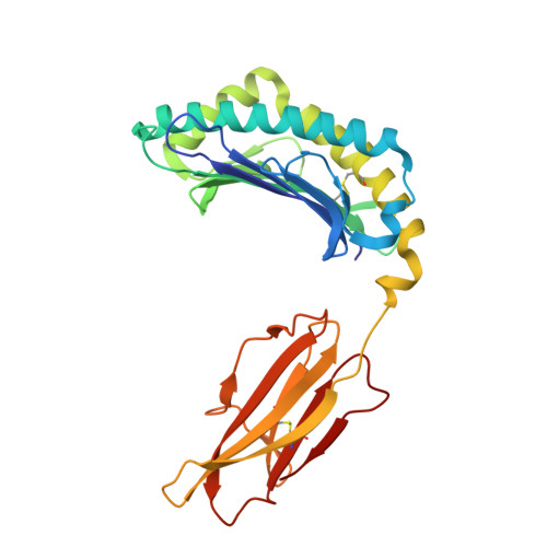
Crystal structure of H-2Db with C.parvum peptide


Experimental Data Snapshot

  • Method: X-RAY DIFFRACTION
  • Resolution: 2.20 Å
  • R-Value Free: 
    0.277 (Depositor), 0.282 (DCC) 
  • R-Value Work: 
    0.230 (Depositor), 0.232 (DCC) 

Starting Model: experimental
View more details

wwPDB Validation 3D Report Full Report

Validation slider image for 20XK

Ligand Structure Quality Assessment 


This is version 1.0 of the entry. See complete history

Literature

Structural Basis for the Immunological Paradox of a High-Affinity Yet Non-Immunogenic MHC-I Epitope from Cryptosporidium parvum.

Fan, S.Wang, T.Ren, S.Peng, J.Li, L.Zhao, Y.Yang, J.Zhang, Y.Yan, Y.Wang, H.Wang, Y.

(2026) Biology (Basel) 15

  • DOI: https://doi.org/10.3390/biology15151281
  • Primary Citation Related Structures: 
    20XK

  • PubMed Abstract: 

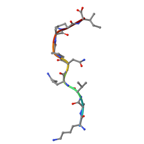
    Cryptosporidium parvum is an important apicomplexan parasite that causes severe diarrheal disease in children and immunocompromised individuals. However, the structural basis for the limited immunogenicity of its T-cell epitopes remains poorly understood. This study integrates structural biology and immunological approaches to elucidate the molecular basis underlying the non-immunogenicity of KAV9, a Cp23-derived epitope with the sequence KAVKNPAPI. Biophysical analyses demonstrated that KAV9 forms a high-affinity complex with H-2D b , with an IC 50 of 7.83 nM, and exhibits higher thermal stability ( T m = 59.16 °C) than the immunodominant LCMV gp33 epitope ( T m = 51.15 °C). Despite strong pMHC binding and high pMHC stability, in vivo peptide immunization failed to elicit a detectable KAV9-specific CD8 + T-cell response. Crystal structure analysis revealed that KAV9 is tightly accommodated within the H-2D b binding groove through an extensive hydrogen-bond network. However, its distinct peptide conformation, particularly involving P4-Lys and the proline residues at P6 and P8, markedly reshapes the TCR-exposed surface compared to gp33. AlphaFold3 (AF3) modeling further suggested that these structural deviations disrupt critical hydrogen-bond interactions with the T-cell receptor (TCR) CDR3 loops, thereby eliminating contacts required for TCRβ engagement. Sequence analysis revealed that KAV9 is highly conserved across multiple Cryptosporidium species, suggesting a conserved structural feature associated with limited T-cell recognition. Together, these findings demonstrate that strong MHC binding and pMHC stability are insufficient to ensure CD8 + T-cell immunogenicity. Instead, the topology of the TCR-accessible peptide surface represents a critical determinant of epitope immunogenicity, with significant implications for epitope selection and vaccine design against cryptosporidiosis.


  • Organizational Affiliation
    • College of Life Sciences and Agronomy, Zhoukou Normal University, Zhoukou 466001, China.

Macromolecule Content 

  • Total Structure Weight: 44.57 kDa 
  • Atom Count: 3,184 
  • Modeled Residue Count: 380 
  • Deposited Residue Count: 382 
  • Unique protein chains: 3

Macromolecules

Find similar proteins by:|  3D Structure
Entity ID: 1
MoleculeChains  Sequence LengthOrganismDetailsImage
H-2 class I histocompatibility antigen, D-B alpha chain275Mus musculusMutation(s): 0 
Gene Names: H2-D1
UniProt
Find proteins for P01899 (Mus musculus)
Explore P01899 
Go to UniProtKB:  P01899
Entity Groups
Sequence Clusters30% Identity50% Identity70% Identity90% Identity95% Identity100% Identity
UniProt GroupP01899
Sequence Annotations
Expand
Reference Sequence
Find similar proteins by:|  3D Structure
Entity ID: 2
MoleculeChains  Sequence LengthOrganismDetailsImage
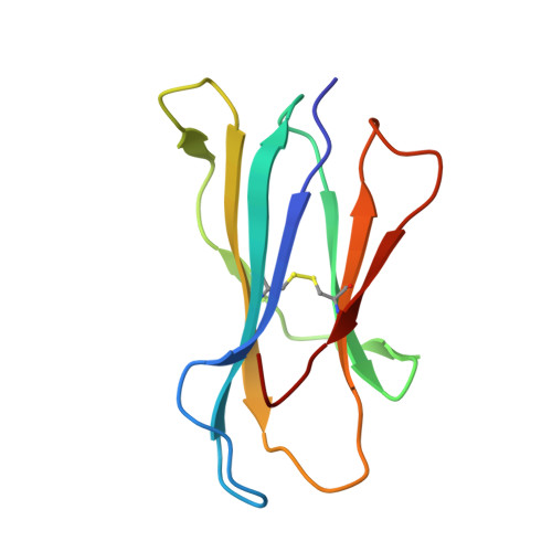
Beta-2-microglobulin98Mus musculusMutation(s): 0 
Gene Names: B2m
UniProt & NIH Common Fund Data Resources
Find proteins for P01887 (Mus musculus)
Explore P01887 
Go to UniProtKB:  P01887
IMPC:  MGI:88127
Entity Groups
Sequence Clusters30% Identity50% Identity70% Identity90% Identity95% Identity100% Identity
UniProt GroupP01887
Sequence Annotations
Expand
Reference Sequence
Find similar proteins by:  Sequence   |   3D Structure  
Entity ID: 3
MoleculeChains  Sequence LengthOrganismDetailsImage
LYS-ALA-VAL-LYS-ASN-PRO-ALA-PRO-ILE9Cryptosporidium parvumMutation(s): 0 
Sequence Annotations
Expand
Reference Sequence

Small Molecules

Ligands 1 Unique
IDChains Name / Formula / InChI Key2D Diagram3D Interactions
GOL
(Subject of Investigation/LOI)

Query on GOL


Download:Ideal Coordinates CCD File
D [auth A]GLYCEROL
C3 H8 O3
PEDCQBHIVMGVHV-UHFFFAOYSA-N

Experimental Data & Validation

Experimental Data

  • Method: X-RAY DIFFRACTION
  • Resolution: 2.20 Å
  • R-Value Free:  0.277 (Depositor), 0.282 (DCC) 
  • R-Value Work:  0.230 (Depositor), 0.232 (DCC) 
Space Group: C 1 2 1
Unit Cell:
Length ( Å )Angle ( ˚ )
a = 91.7α = 90
b = 111.11β = 122.253
c = 58.04γ = 90
Software Package:
Software NamePurpose
REFMACrefinement
HKL-3000data reduction
HKL-3000data scaling
PHASESphasing

Structure Validation

View Full Validation Report


Ligand Structure Quality Assessment 


Entry History 

& Funding Information

Deposition Data


Funding OrganizationLocationGrant Number
National Natural Science Foundation of China (NSFC)China31702232

Revision History  (Full details and data files)

  • Version 1.0: 2026-09-16
    Type: Initial release