1V2I

Structure of the hemagglutinin-neuraminidase from human parainfluenza virus type III


Experimental Data Snapshot

  • Method: X-RAY DIFFRACTION
  • Resolution: 2.20 Å
  • R-Value Free: 0.259 
  • R-Value Work: 0.185 
  • R-Value Observed: 0.185 

wwPDB Validation   3D Report Full Report


Ligand Structure Quality Assessment 


This is version 2.1 of the entry. See complete history


Literature

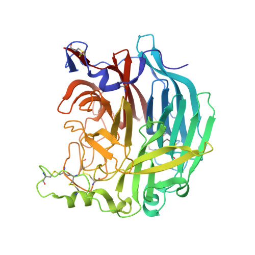
Structure of the Haemagglutinin-neuraminidase from Human Parainfluenza Virus Type III

Lawrence, M.C.Borg, N.A.Streltsov, V.A.Pilling, P.A.Epa, V.C.Varghese, J.N.McKimm-Breschkin, J.L.Colman, P.M.

(2004) J Mol Biol 335: 1343-1357

  • DOI: https://doi.org/10.1016/j.jmb.2003.11.032
  • Primary Citation of Related Structures:  
    1V2I, 1V3B, 1V3C, 1V3D, 1V3E

  • PubMed Abstract: 

    The three-dimensional structure of the haemagglutinin-neuraminidase (HN) from a human parainfluenza virus is described at ca 2.0 A resolution, both in native form and in complex with three substrate analogues. In support of earlier work on the structure of the homologous protein from the avian pathogen Newcastle disease virus (NDV), we observe a dimer of beta-propellers and find no evidence for spatially separated sites performing the receptor-binding and neuraminidase functions of the protein. As with the NDV HN, the active site of the HN of parainfluenza viruses is structurally flexible, suggesting that it may be able to switch between a receptor-binding state and a catalytic state. However, in contrast to the NDV structures, we observe no ligand-induced structural changes that extend beyond the active site and modify the dimer interface.


  • Organizational Affiliation

    CSIRO Health Sciences and Nutrition, 343 Royal Parade, Parkville, Vic 3052, Australia. mike.lawrence@csiro.au


Macromolecules
Find similar proteins by:  (by identity cutoff)  |  3D Structure
Entity ID: 1
MoleculeChains Sequence LengthOrganismDetailsImage
hemagglutinin-neuraminidase glycoprotein
A, B
431Human respirovirus 3Mutation(s): 0 
EC: 3.2.1.18
UniProt
Find proteins for Q6WJ03 (Human respirovirus 3)
Explore Q6WJ03 
Go to UniProtKB:  Q6WJ03
Entity Groups  
Sequence Clusters30% Identity50% Identity70% Identity90% Identity95% Identity100% Identity
UniProt GroupQ6WJ03
Sequence Annotations
Expand
  • Reference Sequence
Oligosaccharides

Help

Entity ID: 2
MoleculeChains Length2D Diagram Glycosylation3D Interactions
alpha-D-mannopyranose-(1-3)-[alpha-D-mannopyranose-(1-6)]beta-D-mannopyranose-(1-4)-2-acetamido-2-deoxy-beta-D-glucopyranose-(1-4)-2-acetamido-2-deoxy-beta-D-glucopyranose
C
5N-Glycosylation
Glycosylation Resources
GlyTouCan:  G22768VO
GlyCosmos:  G22768VO
GlyGen:  G22768VO
Entity ID: 3
MoleculeChains Length2D Diagram Glycosylation3D Interactions
2-acetamido-2-deoxy-beta-D-glucopyranose-(1-4)-2-acetamido-2-deoxy-beta-D-glucopyranose
D, E
2N-Glycosylation
Glycosylation Resources
GlyTouCan:  G42666HT
GlyCosmos:  G42666HT
GlyGen:  G42666HT
Experimental Data & Validation

Experimental Data

  • Method: X-RAY DIFFRACTION
  • Resolution: 2.20 Å
  • R-Value Free: 0.259 
  • R-Value Work: 0.185 
  • R-Value Observed: 0.185 
  • Space Group: P 21 21 21
Unit Cell:
Length ( Å )Angle ( ˚ )
a = 83.5α = 90
b = 93.78β = 90
c = 105.2γ = 90
Software Package:
Software NamePurpose
CNSrefinement
DENZOdata reduction
SCALEPACKdata scaling
X-PLORphasing

Structure Validation

View Full Validation Report


Ligand Structure Quality Assessment 


Entry History 

Deposition Data

Revision History  (Full details and data files)

  • Version 1.0: 2004-02-03
    Type: Initial release
  • Version 1.1: 2008-04-27
    Changes: Version format compliance
  • Version 1.2: 2011-07-13
    Changes: Non-polymer description, Version format compliance
  • Version 2.0: 2020-07-29
    Type: Remediation
    Reason: Carbohydrate remediation
    Changes: Advisory, Atomic model, Data collection, Derived calculations, Non-polymer description, Structure summary
  • Version 2.1: 2023-10-25
    Changes: Data collection, Database references, Refinement description, Structure summary