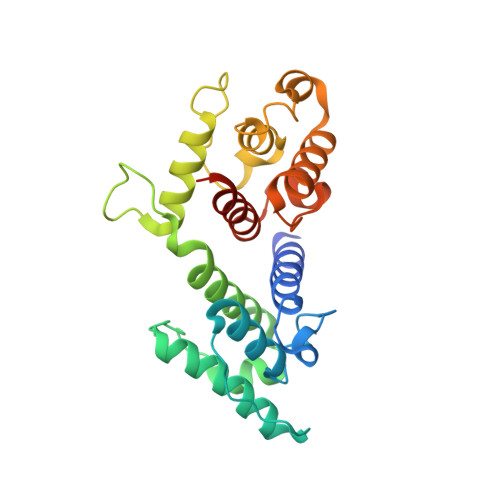
Actin-binding domain of mouse plectin: crystal structure and binding to vimentin
Sevcik, J., Urbanikova, L., Kostan, J., Janda, L., Wiche, G.(2004) Eur J Biochem 271: 1873-1884
- PubMed: 15128297
- DOI: https://doi.org/10.1111/j.1432-1033.2004.04095.x
- Primary Citation of Related Structures:
1SH5, 1SH6 - PubMed Abstract:
Plectin, a large and widely expressed cytolinker protein, is composed of several subdomains that harbor binding sites for a variety of different interaction partners. A canonical actin-binding domain (ABD) comprising two calponin homology domains (CH1 and CH2) is located in proximity to its amino terminus. However, the ABD of plectin is unique among actin-binding proteins as it is expressed in the form of distinct, plectin isoform-specific versions. We have determined the three-dimensional structure of two distinct crystalline forms of one of its ABD versions (pleABD/2alpha) from mouse, to a resolution of 1.95 and 2.0 A. Comparison of pleABD/2alpha with the ABDs of fimbrin and utrophin revealed structural similarity between plectin and fimbrin, although the proteins share only low sequence identity. In fact, pleABD/2alpha has been found to have the same compact fold as the human plectin ABD and the fimbrin ABD, differing from the open conformation described for the ABDs of utrophin and dystrophin. Plectin harbors a specific binding site for intermediate filaments of various types within its carboxy-terminal R5 repeat domain. Our experiments revealed an additional vimentin-binding site of plectin, residing within the CH1 subdomain of its ABD. We show that vimentin binds to this site via the amino-terminal part of its rod domain. This additional amino-terminal intermediate filament protein binding site of plectin may have a function in intermediate filament dynamics and assembly, rather than in linking and stabilizing intermediate filament networks.
- Institute of Molecular Biology, Slovak Academy of Sciences, Bratislava, Slovak Republic.
Organizational Affiliation: