NMR Solution Structure of the Focal Adhesion Targeting Domain of Focal Adhesion Kinase in Complex with a Paxillin LD Peptide: EVIDENCE FOR A TWO-SITE BINDING MODEL.
Gao, G., Prutzman, K.C., King, M.L., Scheswohl, D.M., DeRose, E.F., London, R.E., Schaller, M.D., Campbell, S.L.(2004) J Biological Chem 279: 8441-8451
- PubMed: 14662767
- DOI: https://doi.org/10.1074/jbc.M309808200
- Primary Citation of Related Structures:
1QVX - PubMed Abstract:
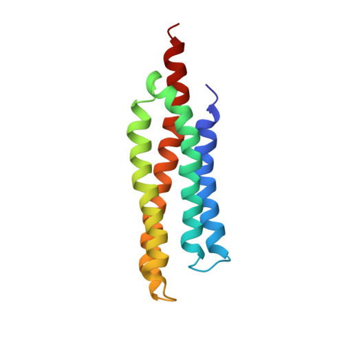
Focal adhesion kinase (FAK) is a non-receptor tyrosine kinase that is regulated by integrins. Upon activation, FAK generates signals that modulate crucial cell functions, including cell proliferation, migration, and survival. The C-terminal focal adhesion targeting (FAT) sequence mediates localization of FAK to discrete regions in the cell called focal adhesions. Several binding partners for the FAT domain of FAK have been identified, including paxillin. We have determined the solution structure of the avian FAT domain in complex with a peptide mimicking the LD2 motif of paxillin by NMR spectroscopy. The FAT domain retains a similar fold to that found in the unliganded form when complexed to the paxillin-derived LD2 peptide, an antiparallel four-helix bundle. However, noticeable conformational changes were observed upon the LD2 peptide binding, especially the position of helix 4. Multiple lines of evidence, including the results obtained from isothermal titration calorimetry, intermolecular nuclear Overhauser effects, mutagenesis, and protection from paramagnetic line broadening, support the existence of two distinct paxillin-binding sites on the opposite faces of the FAT domain. The structure of the FAT domain-LD2 complex was modeled using the program HADDOCK based on our solution structure of the LD2-bound FAT domain and mutagenesis data. Our model of the FAT domain-LD2 complex provides insight into the molecular basis of FAK-paxillin binding interactions, which will aid in understanding the role of paxillin in FAK targeting and signaling.
- Department of Biochemistry and Biophysics, University of North Carolina, Chapel Hill, North Carolina 27599, USA.
Organizational Affiliation: