1PER

THE COMPLEX BETWEEN PHAGE 434 REPRESSION DNA-BINDING DOMAIN AND OPERATOR SITE OR3: STRUCTURAL DIFFERENCES BETWEEN CONSENSUS AND NON-CONSENSUS HALF-SITES


Experimental Data Snapshot

  • Method: X-RAY DIFFRACTION
  • Resolution: 2.50 Å
  • R-Value Observed: 0.187 

wwPDB Validation   3D Report Full Report


This is version 1.3 of the entry. See complete history


Literature

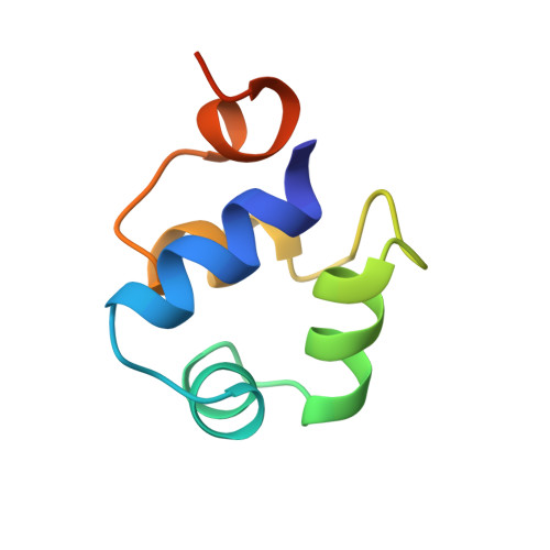
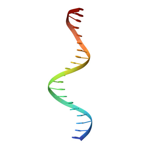
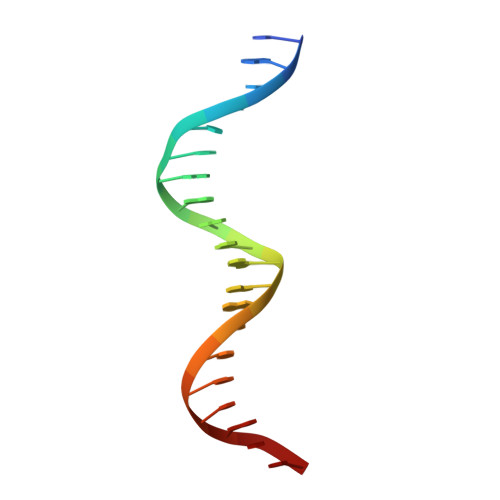
The complex between phage 434 repressor DNA-binding domain and operator site OR3: structural differences between consensus and non-consensus half-sites.

Rodgers, D.W.Harrison, S.C.

(1993) Structure 1: 227-240

  • DOI: https://doi.org/10.1016/0969-2126(93)90012-6
  • Primary Citation of Related Structures:  
    1PER

  • PubMed Abstract: 

    The repressor of phage 434 binds to a set of operator sites as a homodimer. Its relative affinities for these sites determine the switch from lysogenic to lytic growth. The six 434 operator sites (OR1, OR2, OR3, OL1, OL2 and OL3) have a particularly simple organization; all are 14 base pairs long, with a conserved 5'-ACAA sequence symmetrically placed at either end, and a variable central six base pairs. OR3 is unique among naturally-occurring 434 operator sites in that it contains a non-consensus base pair, G.C, at the fourth position of the otherwise invariant 5'-ACAA sequence. Comparisons among structures of the 434 repressor DNA-binding domain, R1-69, bound to various operator sites, allow us to analyze differential specificity in regulatory complexes of this kind.


  • Organizational Affiliation

    Howard Hughes Medical Institute, Harvard University, Cambridge, MA 02138.


Macromolecules

Find similar proteins by:  (by identity cutoff)  |  3D Structure
Entity ID: 3
MoleculeChains Sequence LengthOrganismDetailsImage
PROTEIN (434 REPRESSOR)C [auth L],
D [auth R]
69Phage 434Mutation(s): 0 
UniProt
Find proteins for P16117 (Enterobacteria phage 434)
Explore P16117 
Go to UniProtKB:  P16117
Entity Groups  
Sequence Clusters30% Identity50% Identity70% Identity90% Identity95% Identity100% Identity
UniProt GroupP16117
Sequence Annotations
Expand
  • Reference Sequence

Find similar nucleic acids by:  Sequence   |   3D Structure  

Entity ID: 1
MoleculeChains LengthOrganismImage
DNA (5'-D(*AP*AP*GP*TP*AP*CP*AP*GP*TP*TP*TP*TP*TP*CP*TP*TP*G P*TP*AP*T)-3')20N/A
Sequence Annotations
Expand
  • Reference Sequence

Find similar nucleic acids by:  Sequence   |   3D Structure  

Entity ID: 2
MoleculeChains LengthOrganismImage
DNA (5'-D(*TP*AP*TP*AP*CP*AP*AP*GP*AP*AP*AP*AP*AP*CP*TP*GP*T P*AP*CP*T)-3')20N/A
Sequence Annotations
Expand
  • Reference Sequence
Experimental Data & Validation

Experimental Data

  • Method: X-RAY DIFFRACTION
  • Resolution: 2.50 Å
  • R-Value Observed: 0.187 
  • Space Group: P 21 21 21
Unit Cell:
Length ( Å )Angle ( ˚ )
a = 150.9α = 90
b = 64.5β = 90
c = 27.8γ = 90
Software Package:
Software NamePurpose
CORELSrefinement
TNTrefinement

Structure Validation

View Full Validation Report


Entry History 

Deposition Data

Revision History  (Full details and data files)

  • Version 1.0: 1994-01-31
    Type: Initial release
  • Version 1.1: 2008-05-22
    Changes: Version format compliance
  • Version 1.2: 2011-07-13
    Changes: Version format compliance
  • Version 1.3: 2024-02-14
    Changes: Data collection, Database references