1LX7

Structure of E. coli uridine phosphorylase at 2.0A


Experimental Data Snapshot

  • Method: X-RAY DIFFRACTION
  • Resolution: 2.00 Å
  • R-Value Free: 0.215 
  • R-Value Work: 0.182 

wwPDB Validation   3D Report Full Report


This is version 1.3 of the entry. See complete history


Literature

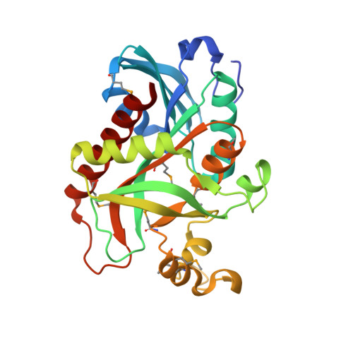
Structure of Escherichia coli uridine phosphorylase at 2.0 A.

Burling, F.T.Kniewel, R.Buglino, J.A.Chadha, T.Beckwith, A.Lima, C.D.

(2003) Acta Crystallogr D Biol Crystallogr 59: 73-76

  • DOI: https://doi.org/10.1107/s0907444902018929
  • Primary Citation of Related Structures:  
    1LX7

  • PubMed Abstract: 

    The 2.0 A crystal structure has been determined for Escherichia coli uridine phosphorylase (UP), an essential enzyme in nucleotide biosynthesis that catalyzes the phosphorolytic cleavage of the C-N glycosidic bond of uridine to ribose-1-phosphate and uracil. The structure determination of two independent monomers in the asymmetric unit revealed the residue composition and atomic details of the apo configurations of each active site. The native hexameric UP enzyme was revealed by applying threefold crystallographic symmetry to the contents of the asymmetric unit. The 2.0 A model reveals a closer structural relationship to other nucleotide phosphorylase enzymes than was previously appreciated.


  • Organizational Affiliation

    Biochemistry Department and Structural Biology Program, Weill Medical College of Cornell University, New York, NY 10021, USA.


Macromolecules
Find similar proteins by:  (by identity cutoff)  |  3D Structure
Entity ID: 1
MoleculeChains Sequence LengthOrganismDetailsImage
uridine phosphorylase
A, B
253Escherichia coliMutation(s): 8 
EC: 2.4.2.3
UniProt
Find proteins for P12758 (Escherichia coli (strain K12))
Explore P12758 
Go to UniProtKB:  P12758
Entity Groups  
Sequence Clusters30% Identity50% Identity70% Identity90% Identity95% Identity100% Identity
UniProt GroupP12758
Sequence Annotations
Expand
  • Reference Sequence
Small Molecules
Modified Residues  1 Unique
IDChains TypeFormula2D DiagramParent
MSE
Query on MSE
A, B
L-PEPTIDE LINKINGC5 H11 N O2 SeMET
Experimental Data & Validation

Experimental Data

  • Method: X-RAY DIFFRACTION
  • Resolution: 2.00 Å
  • R-Value Free: 0.215 
  • R-Value Work: 0.182 
  • Space Group: H 3
Unit Cell:
Length ( Å )Angle ( ˚ )
a = 151.375α = 90
b = 151.375β = 90
c = 48.151γ = 120
Software Package:
Software NamePurpose
DENZOdata reduction
SCALEPACKdata scaling
SOLVEphasing
CNSrefinement

Structure Validation

View Full Validation Report


Entry History 

Revision History  (Full details and data files)

  • Version 1.0: 2002-06-12
    Type: Initial release
  • Version 1.1: 2008-04-28
    Changes: Version format compliance
  • Version 1.2: 2011-07-13
    Changes: Derived calculations, Version format compliance
  • Version 1.3: 2021-02-03
    Changes: Database references, Derived calculations, Structure summary