1LKF

LEUKOCIDIN F (HLGB) FROM STAPHYLOCOCCUS AUREUS


Experimental Data Snapshot

  • Method: X-RAY DIFFRACTION
  • Resolution: 1.90 Å
  • R-Value Free: 0.282 
  • R-Value Work: 0.196 
  • R-Value Observed: 0.196 

wwPDB Validation   3D Report Full Report


This is version 1.3 of the entry. See complete history


Literature

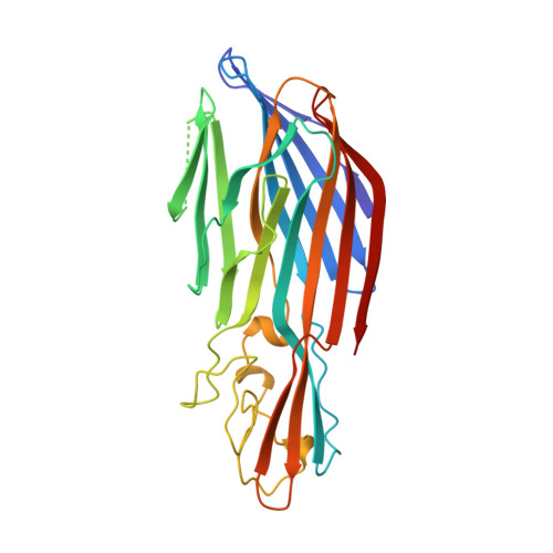
Crystal structure of staphylococcal LukF delineates conformational changes accompanying formation of a transmembrane channel.

Olson, R.Nariya, H.Yokota, K.Kamio, Y.Gouaux, E.

(1999) Nat Struct Biol 6: 134-140

  • DOI: https://doi.org/10.1038/5821
  • Primary Citation of Related Structures:  
    1LKF, 2LKF, 3LKF

  • PubMed Abstract: 

    Staphylococcal LukF, LukS, HgammaII, and alpha-hemolysin are self-assembling, channel-forming proteins related in sequence and function. In the alpha-hemolysin heptamer, the channel-forming beta-strands and the amino latch make long excursions from the protomer core. Here we report the crystal structure of the water soluble form of LukF. In the LukF structure the channel-forming region folds into an amphipathic, three-strand beta-sheet and the amino latch forms a beta-strand extending a central beta-sheet. The LukF structure illustrates how a channel-forming toxin masks protein-protein and protein-membrane interfaces prior to cell binding and assembly, and together with the alpha-hemolysin heptamer structure, they define the end points on the pathway of toxin assembly.


  • Organizational Affiliation

    Department of Biochemistry and Molecular Biophysics, Columbia University, New York, New York 10032, USA.


Macromolecules
Find similar proteins by:  (by identity cutoff)  |  3D Structure
Entity ID: 1
MoleculeChains Sequence LengthOrganismDetailsImage
LEUKOCIDIN F SUBUNIT299Staphylococcus aureusMutation(s): 0 
Gene Names: LUKF
Membrane Entity: Yes 
UniProt
Find proteins for P0A077 (Staphylococcus aureus)
Explore P0A077 
Go to UniProtKB:  P0A077
Entity Groups  
Sequence Clusters30% Identity50% Identity70% Identity90% Identity95% Identity100% Identity
UniProt GroupP0A077
Sequence Annotations
Expand
  • Reference Sequence
Experimental Data & Validation

Experimental Data

  • Method: X-RAY DIFFRACTION
  • Resolution: 1.90 Å
  • R-Value Free: 0.282 
  • R-Value Work: 0.196 
  • R-Value Observed: 0.196 
  • Space Group: P 21 21 21
Unit Cell:
Length ( Å )Angle ( ˚ )
a = 39.76α = 90
b = 48.53β = 90
c = 147.51γ = 90
Software Package:
Software NamePurpose
SOLVEphasing
X-PLORmodel building
X-PLORrefinement
DENZOdata reduction
SCALEPACKdata scaling
X-PLORphasing

Structure Validation

View Full Validation Report


Entry History 

Deposition Data

Revision History  (Full details and data files)

  • Version 1.0: 1999-07-28
    Type: Initial release
  • Version 1.1: 2008-03-24
    Changes: Version format compliance
  • Version 1.2: 2011-07-13
    Changes: Version format compliance
  • Version 1.3: 2024-02-14
    Changes: Data collection, Database references