1KMQ

Crystal Structure of a Constitutively Activated RhoA Mutant (Q63L)


Experimental Data Snapshot

  • Method: X-RAY DIFFRACTION
  • Resolution: 1.55 Å
  • R-Value Free: 0.190 
  • R-Value Work: 0.161 
  • R-Value Observed: 0.163 

wwPDB Validation   3D Report Full Report


Ligand Structure Quality Assessment 


This is version 1.4 of the entry. See complete history


Literature

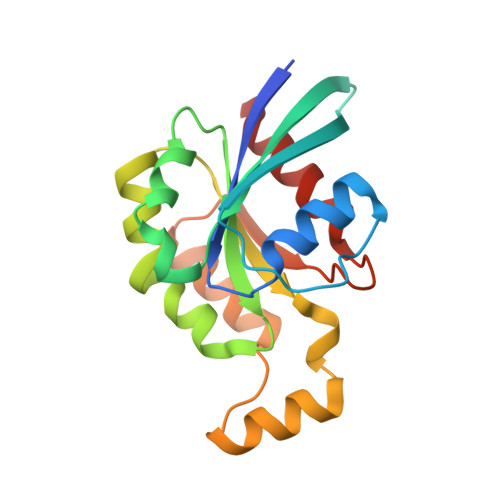
Structure of a constitutively activated RhoA mutant (Q63L) at 1.55 A resolution.

Longenecker, K.Read, P.Lin, S.K.Somlyo, A.P.Nakamoto, R.K.Derewenda, Z.S.

(2003) Acta Crystallogr D Biol Crystallogr 59: 876-880

  • DOI: https://doi.org/10.1107/s0907444903005390
  • Primary Citation of Related Structures:  
    1KMQ

  • PubMed Abstract: 

    Mutants of the small G protein RhoA that are deficient in GTPase activity and thereby exhibit constitutive molecular signaling activity are commonly used to discover its cellular functions. In particular, two such mutants, Gly14-->Val (G14V) and Gln63-->Leu (Q63L), are often used interchangeably for such studies. However, while their in vitro rates of GTP hydrolysis are very similar, differences are observed in their other functional properties. The structure of G14V-RhoA is known; in order to assess whether structural variations are responsible for functional differences, the crystal structure of a Q63L-RhoA bound to the GTP-analog 5'-guanylylimidodiphosphate (GMPPNP) was determined at 1.5 A resolution. Overall, the structure is very similar to that of G14V-RhoA, but the significantly higher resolution data permit an improved basis for structural analysis and comparison. The data support the notion that differences observed between the mutants in vivo are likely to arise from altered affinities for RhoGDI and not from direct structural differences.


  • Organizational Affiliation

    Department of Molecular Physiology and Biological Physics, University of Virginia, Charlottesville, VA 22908, USA.


Macromolecules
Find similar proteins by:  (by identity cutoff)  |  3D Structure
Entity ID: 1
MoleculeChains Sequence LengthOrganismDetailsImage
TRANSFORMING PROTEIN RHOA184Homo sapiensMutation(s): 2 
UniProt & NIH Common Fund Data Resources
Find proteins for P61586 (Homo sapiens)
Explore P61586 
Go to UniProtKB:  P61586
PHAROS:  P61586
GTEx:  ENSG00000067560 
Entity Groups  
Sequence Clusters30% Identity50% Identity70% Identity90% Identity95% Identity100% Identity
UniProt GroupP61586
Sequence Annotations
Expand
  • Reference Sequence
Experimental Data & Validation

Experimental Data

  • Method: X-RAY DIFFRACTION
  • Resolution: 1.55 Å
  • R-Value Free: 0.190 
  • R-Value Work: 0.161 
  • R-Value Observed: 0.163 
  • Space Group: P 21 21 2
Unit Cell:
Length ( Å )Angle ( ˚ )
a = 60.581α = 90
b = 73.34β = 90
c = 48.041γ = 90
Software Package:
Software NamePurpose
DENZOdata reduction
SCALEPACKdata scaling
AMoREphasing
REFMACrefinement

Structure Validation

View Full Validation Report


Ligand Structure Quality Assessment 


Entry History 

Deposition Data

Revision History  (Full details and data files)

  • Version 1.0: 2003-05-06
    Type: Initial release
  • Version 1.1: 2008-04-27
    Changes: Version format compliance
  • Version 1.2: 2011-07-13
    Changes: Version format compliance
  • Version 1.3: 2021-10-27
    Changes: Database references, Derived calculations
  • Version 1.4: 2024-02-14
    Changes: Data collection